adnan923060792027@gmail.com

A visual comparison of original, reconstructed, and noise-injected medical images under federated learning to illustrate privacy risks and shadow defense impact.

🔒7 Alarming Privacy Risks of Federated Learning—and the Breakthrough Shadow Defense Fix You Need

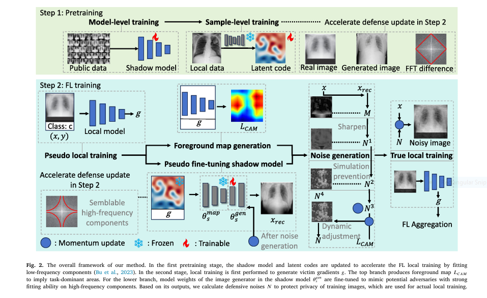
Introduction Federated Learning (FL) has been heralded as the privacy-preserving future of AI, especially in sensitive domains like healthcare. But behind its collaborative promise lies a serious vulnerability: gradient inversion attacks (GIA). These attacks can reconstruct original training images from shared gradients—exposing confidential patient data. Enter the breakthrough: Shadow Defense. In this article, we dive […]

🔒7 Alarming Privacy Risks of Federated Learning—and the Breakthrough Shadow Defense Fix You Need Read More »

Simulation of individualized brain aging and Alzheimer’s progression using AI with diffeomorphic registration

🧠 7 Groundbreaking Insights from a Revolutionary Brain Aging AI Model You Can’t Ignore

Introduction Predicting the trajectory of brain aging—whether due to normal aging or the onset of Alzheimer’s Disease (AD)—has always posed a massive challenge. But what if you could simulate future brain scans using just a single MRI? That’s exactly what the new InBrainSyn framework achieves using deep generative models and parallel transport. In this article,

🧠 7 Groundbreaking Insights from a Revolutionary Brain Aging AI Model You Can’t Ignore Read More »

Disentangled generative model showcasing independent factors of age, ethnicity, and camera in synthetic retinal images

🔍 7 Breakthrough Insights: How Disentangled Generative Models Fix Biases in Retinal Imaging (and Where They Fail)

Introduction: Why Bias in Retinal Imaging Matters More Than Ever Retinal fundus images are crucial in diagnosing conditions from diabetic retinopathy to cardiovascular diseases. But here’s the problem: most AI models trained on retinal images learn the wrong things. Imagine this: a deep learning system that diagnoses ethnicity instead of actual disease features—because the camera

🔍 7 Breakthrough Insights: How Disentangled Generative Models Fix Biases in Retinal Imaging (and Where They Fail) Read More »

Medical AI transforming tumor segmentation with EGTA-KD technology

Revolutionary AI Breakthrough: Non-Contrast Tumor Segmentation Saves Lives & Avoids Deadly Risks

Imagine detecting deadly tumors without injecting risky contrast agents. A revolutionary AI framework called EGTA-KD is making this possible, achieving near-perfect segmentation (90.8% accuracy) on non-contrast scans while eliminating allergic reactions and kidney damage linked to traditional methods. This isn’t futuristic hype – it’s validated across brain, liver, and kidney tumors in major clinical datasets. The Deadly Cost of Current

Revolutionary AI Breakthrough: Non-Contrast Tumor Segmentation Saves Lives & Avoids Deadly Risks Read More »

Counterfactual contrastive learning closes the performance gap between majority and minority scanners, especially where it matters most: in low-data settings and real-world deployments

Title: 5 Powerful Reasons Why Counterfactual Contrastive Learning Beats Traditional Medical Imaging Techniques (And How It Can Transform Your Practice)

Introduction: The Future of Medical Imaging Starts Here Medical imaging has long been a cornerstone of diagnostics, but traditional methods often fall short when it comes to adapting to real-world variability. Enter counterfactual contrastive learning , an innovative framework that’s changing the game by leveraging causal image synthesis to improve model robustness and downstream performance.

Title: 5 Powerful Reasons Why Counterfactual Contrastive Learning Beats Traditional Medical Imaging Techniques (And How It Can Transform Your Practice) Read More »

RO-LMM AI system seamlessly processing MRI, ultrasound, and pathology reports to generate precise radiotherapy plans and 3D target segmentations for a breast cancer patient."

Beyond Human Limits 1: How RO-LMM’s AI is Revolutionizing Breast Cancer Radiotherapy Planning (Saving Lives & Time)

The Crippling Burden of Breast Cancer Radiotherapy Planning (And the AI Solution Changing Everything) Every 38 seconds, a woman is diagnosed with breast cancer globally. For these patients, timely and precise radiotherapy is often a lifeline. Yet, the complex, multi-step process of planning this treatment – involving synthesizing medical reports, defining treatment strategies, and meticulously mapping

Beyond Human Limits 1: How RO-LMM’s AI is Revolutionizing Breast Cancer Radiotherapy Planning (Saving Lives & Time) Read More »

SVIS-RULEX SFMOV heatmap overlay on a chest X-ray: Red/Orange areas highlight regions of high statistical significance (e.g., mean intensity, skewness, entropy) corresponding to COVID-19 lung opacities, validated by radiologists. Blue areas show less relevant tissue

3 Breakthroughs & 1 Warning: How Explainable AI SVIS-RULEX is Revolutionizing Medical Imaging (Finally!)

For years, artificial intelligence (AI) has promised to revolutionize medical diagnosis, particularly in analyzing complex medical images like X-rays, MRIs, and ultrasounds. Deep learning models consistently achieve superhuman accuracy in spotting tumors, infections, and subtle pathologies. Yet, a critical roadblock remains: the “black box” problem. How does the AI really make its decision? Without transparency, doctors hesitate to

3 Breakthroughs & 1 Warning: How Explainable AI SVIS-RULEX is Revolutionizing Medical Imaging (Finally!) Read More »

ElastoNet: A revolutionary neural network approach to MR Elastography inversion with uncertainty quantification.

ElastoNet 1: The Revolutionary Neural Network for MRE Wave Inversion with Uncertainty Quantification (Pros & Cons)

Introduction: Why ElastoNet Is Changing the Game in Medical Imaging Medical imaging has seen a rapid evolution over the past decade, especially in non-invasive diagnostics. Among these advancements, Magnetic Resonance Elastography (MRE) has emerged as a powerful technique for evaluating tissue stiffness — a key biomarker in diagnosing diseases like liver fibrosis and cancer. However,

ElastoNet 1: The Revolutionary Neural Network for MRE Wave Inversion with Uncertainty Quantification (Pros & Cons) Read More »

AI algorithms analyzing 3D TOF-MRA scans of the Circle of Willis for aneurysm risk prediction in Crown challenge

CROWN Challenge Breakthrough: 6 AI Solutions Transform Brain Artery Analysis (But Still Fall Short)

Why Intracranial Aneurysm Screening Is Failing Patients Intracranial aneurysms (IAs) affect 3% of the global population, yet rupture often strikes without warning. The CROWN Challenge—a landmark MICCAI 2023 study—reveals a critical gap: current IA screening misses 92% of at-risk cases. Traditional manual assessment of the Circle of Willis (CoW) is slow, inconsistent, and fails to leverage key

CROWN Challenge Breakthrough: 6 AI Solutions Transform Brain Artery Analysis (But Still Fall Short) Read More »

Advanced 3D visualization of a cardiology digital twin showing ventricular activation and ECG mapping

7 Groundbreaking Innovations in Cardiac Digital Twins: Unlocking the Future of Precision Cardiology (and 3 Major Challenges Holding It Back)

The Non-Invasive Revolution in Cardiac Mapping Imagine holding a perfect digital replica of your heart that beats like the real thing, predicts how you’ll respond to treatments, and pinpoints electrical flaws without invasive tests. This isn’t science fiction—it’s the promise of Cardiac Digital Twins (CDTs), and a groundbreaking study just cracked a critical code: decoding your heart’s hidden

7 Groundbreaking Innovations in Cardiac Digital Twins: Unlocking the Future of Precision Cardiology (and 3 Major Challenges Holding It Back) Read More »

Follow by Email
Tiktok