AI in medical imaging

Revolutionizing Medical Image Segmentation: SemSim’s Semantic Breakthrough

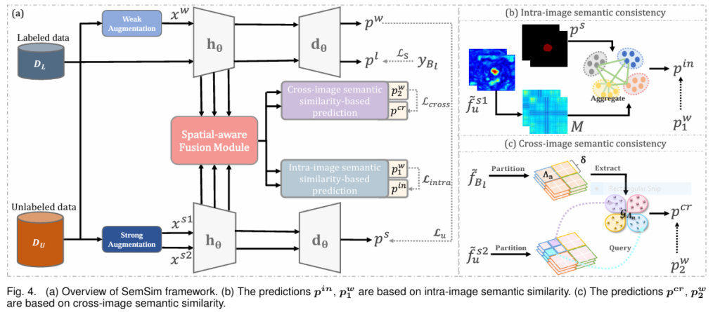
Medical image segmentation is the cornerstone of modern diagnostics and treatment planning. From pinpointing tumor boundaries to mapping cardiac structures, its precision directly impacts patient outcomes. Yet, a critical bottleneck persists: the massive annotation burden. Manual labeling demands hours of expert time per scan, creating a severe shortage of labeled data that throttles AI’s potential. Enter semi-supervised learning […]

Revolutionizing Medical Image Segmentation: SemSim’s Semantic Breakthrough Read More »

Complete overview of proposed GGLA-NeXtE2NET network.

GGLA-NeXtE2NET: Advanced Brain Tumor Recognition

The accurate and timely diagnosis of brain tumors is a critical challenge in modern medicine. Magnetic Resonance Imaging (MRI) is an essential non-invasive tool that provides detailed images of the brain’s internal structures, helping to identify the size, location, and type of tumors. However, the interpretation of these images can be complex due to the

GGLA-NeXtE2NET: Advanced Brain Tumor Recognition Read More »

Architecture of DGLA-ResNet50 model. (a) Structure of GLA Bneck feature extraction module.

Enhancing Skin Lesion Detection Accuracy

Skin cancer continues to be one of the fastest-growing cancers worldwide, with early detection being critical for effective treatment. Traditional diagnostic methods rely heavily on dermatologists’ expertise and dermoscopy, a non-invasive skin imaging technique. However, the manual nature of dermoscopy makes the process time-consuming and subjective. To overcome these limitations, the research paper titled “Skin

Enhancing Skin Lesion Detection Accuracy Read More »

The proposed multi-label skin lesion classification framework has three branches: dermoscopy imaging modality branch (green block), clinical imaging modality branch (yellow block), and a hybrid-meta branch (orange block). Modified Xception module based dermoscopy and clinical imaging modalities’ features are first concatenated, then fed to the input of hybrid-meta branch, and finally concatenated with the meta-data.

AI Revolutionizes Skin Cancer Diagnosis

New Deep Learning Model Boosts Accuracy for Early Detection Skin cancer, particularly melanoma, remains one of the deadliest cancers worldwide. The stakes for early detection couldn’t be higher: diagnose melanoma at an advanced stage, and the 10-year survival rate plummets to a grim 39%. Catch it early, however, and survival rates soar above 93%. This

AI Revolutionizes Skin Cancer Diagnosis Read More »

Schematic representation of the proposed LungCT-NET, incorporating preprocessing, reconstructed transfer learning (TL) models, stacking ensemble learning, SHAP (Shapley Additive Explanations) for explainable artificial intelligence (XAI), along with model evaluation and comparison.

LungCT-NET: Revolutionizing Lung Cancer Diagnosis with AI

Introduction: The Urgent Need for Early Lung Cancer Detection Lung cancer is the leading cause of cancer-related deaths worldwide, accounting for 1.8 million fatalities in 2020 alone. Its deadliness is largely due to late diagnosis, as early-stage symptoms are often indistinct. Detecting malignant lung nodules from CT scans early can significantly improve survival rates. However,

LungCT-NET: Revolutionizing Lung Cancer Diagnosis with AI Read More »

Brain Tumor Diagnosis: GATE-CNN

Revolutionizing Brain Tumor Diagnosis: GATE-CNN

For patients facing a potential brain tumor diagnosis, time is brain tissue. Early and accurate detection isn’t just beneficial; it’s often the solitary lifeline separating treatable conditions from devastating outcomes. Magnetic Resonance Imaging (MRI) stands as the cornerstone of brain tumor visualization, offering unparalleled detail of the brain’s intricate structures. Yet, interpreting these complex images

Revolutionizing Brain Tumor Diagnosis: GATE-CNN Read More »

Fig. 1. The overall framework of our multi-teacher distillation method.

Adaptive Multi-Teacher Knowledge Distillation for Segmentation

Medical image segmentation is a cornerstone of modern diagnostics, enabling precise identification of tumors, organs, and anomalies in MRI and CT scans. However, challenges like limited data, privacy concerns, and the computational complexity of deep learning models hinder their real-world adoption. Enter adaptive multi-teacher knowledge distillation—a groundbreaking approach that balances accuracy, efficiency, and privacy. In this

Adaptive Multi-Teacher Knowledge Distillation for Segmentation Read More »

3DL-Net’s three-stage architecture: preliminary segmentation, multi-scale context extraction, and dendritic refinement for precise medical image analysis.

Revolutionizing Medical Image Segmentation with 3DL-Net: A Breakthrough in Global–Local Feature Representation

Medical image segmentation is a cornerstone of modern healthcare, enabling precise delineation of anatomical structures and pathological regions. From aiding accurate clinical assessments to facilitating disease diagnosis and treatment planning, its applications span across various imaging modalities such as CT scans, MRIs, and ultrasounds. However, achieving precise and efficient segmentation remains a formidable challenge due

Revolutionizing Medical Image Segmentation with 3DL-Net: A Breakthrough in Global–Local Feature Representation Read More »

Advances in Attention Mechanisms for Medical Image Segmentation: A Comprehensive Guide

Medical image segmentation is a cornerstone of modern healthcare, enabling precise diagnosis and treatment planning through advanced imaging technologies. As deep learning continues to evolve, attention mechanisms have emerged as a game-changer in enhancing the accuracy and efficiency of medical image segmentation. This article delves into the latest advancements in attention mechanisms, drawing insights from

Advances in Attention Mechanisms for Medical Image Segmentation: A Comprehensive Guide Read More »