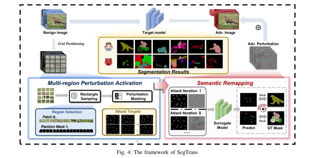
adnan923060792027@gmail.com

Architecture of DGLA-ResNet50 model. (a) Structure of GLA Bneck feature extraction module.

Enhancing Skin Lesion Detection Accuracy

Skin cancer continues to be one of the fastest-growing cancers worldwide, with early detection being critical for effective treatment. Traditional diagnostic methods rely heavily on dermatologists’ expertise and dermoscopy, a non-invasive skin imaging technique. However, the manual nature of dermoscopy makes the process time-consuming and subjective. To overcome these limitations, the research paper titled “Skin […]

Enhancing Skin Lesion Detection Accuracy Read More »

The proposed multi-label skin lesion classification framework has three branches: dermoscopy imaging modality branch (green block), clinical imaging modality branch (yellow block), and a hybrid-meta branch (orange block). Modified Xception module based dermoscopy and clinical imaging modalities’ features are first concatenated, then fed to the input of hybrid-meta branch, and finally concatenated with the meta-data.

AI Revolutionizes Skin Cancer Diagnosis

New Deep Learning Model Boosts Accuracy for Early Detection Skin cancer, particularly melanoma, remains one of the deadliest cancers worldwide. The stakes for early detection couldn’t be higher: diagnose melanoma at an advanced stage, and the 10-year survival rate plummets to a grim 39%. Catch it early, however, and survival rates soar above 93%. This

AI Revolutionizes Skin Cancer Diagnosis Read More »

Illustration of the framework of the proposed method. In the first stage, an adversarial image is processed with multiscale analysis: the image will be downsampled by a factor of 1/2 and 1/4, respectively, and upsampled by a factor of 2. Then in the second stage, we design and insert 𝑁 diffusive and denoising aggregation mechanism (DDA) blocks sequentially. Each DDA block involves a diffusive process (Section 3.2), a denoising process (Section 3.3), and an aggregation process (Section 3.4). The output samples from the last DDA block will be inversely processed to the original scale and smoothed to obtain the reversed image.

Skin Cancer AI Combats Adversarial Attacks with MDDA

In recent years, deep learning has revolutionized dermatology by automating skin cancer diagnosis with impressive accuracy. AI-powered systems like convolutional neural networks (CNNs) can now detect melanoma and other lesions with expert-level precision. However, alongside these advancements arises a critical vulnerability: adversarial attacks. These are subtle, often imperceptible image perturbations that can mislead even the

Skin Cancer AI Combats Adversarial Attacks with MDDA Read More »

Schematic representation of the proposed LungCT-NET, incorporating preprocessing, reconstructed transfer learning (TL) models, stacking ensemble learning, SHAP (Shapley Additive Explanations) for explainable artificial intelligence (XAI), along with model evaluation and comparison.

LungCT-NET: Revolutionizing Lung Cancer Diagnosis with AI

Introduction: The Urgent Need for Early Lung Cancer Detection Lung cancer is the leading cause of cancer-related deaths worldwide, accounting for 1.8 million fatalities in 2020 alone. Its deadliness is largely due to late diagnosis, as early-stage symptoms are often indistinct. Detecting malignant lung nodules from CT scans early can significantly improve survival rates. However,

LungCT-NET: Revolutionizing Lung Cancer Diagnosis with AI Read More »

Brain Tumor Diagnosis: GATE-CNN

Revolutionizing Brain Tumor Diagnosis: GATE-CNN

For patients facing a potential brain tumor diagnosis, time is brain tissue. Early and accurate detection isn’t just beneficial; it’s often the solitary lifeline separating treatable conditions from devastating outcomes. Magnetic Resonance Imaging (MRI) stands as the cornerstone of brain tumor visualization, offering unparalleled detail of the brain’s intricate structures. Yet, interpreting these complex images

Revolutionizing Brain Tumor Diagnosis: GATE-CNN Read More »

The block diagram of the Brain-GCN-Net model for Brain Tumor Diagnosis.

Beyond the Naked Eye: How AI Fusion is Revolutionizing Brain Tumor Diagnosis

Every year, thousands face the daunting diagnosis of a brain tumor. Speed and accuracy are paramount – early detection significantly improves survival rates and treatment outcomes. Yet, interpreting complex MRI scans remains a challenging, time-consuming task for even the most skilled radiologists. Misdiagnosis or delayed diagnosis can have devastating consequences. Enter artificial intelligence (AI), poised

Beyond the Naked Eye: How AI Fusion is Revolutionizing Brain Tumor Diagnosis Read More »

Fig. 1. The overall framework of our multi-teacher distillation method.

Adaptive Multi-Teacher Knowledge Distillation for Segmentation

Medical image segmentation is a cornerstone of modern diagnostics, enabling precise identification of tumors, organs, and anomalies in MRI and CT scans. However, challenges like limited data, privacy concerns, and the computational complexity of deep learning models hinder their real-world adoption. Enter adaptive multi-teacher knowledge distillation—a groundbreaking approach that balances accuracy, efficiency, and privacy. In this

Adaptive Multi-Teacher Knowledge Distillation for Segmentation Read More »

The flowchart of the medical image classification with SAM-based Image Enhancement (SAM-IE). The terms ‘low-grade’ and ‘high-grade’ can refer to benign and malignant, respectively, or to different degrees of disease severity.

SAM-IE: Enhancing Medical Imaging for Disease Detection

Medical imaging is a cornerstone of modern diagnostics, yet clinicians often grapple with challenges like ambiguous anatomical structures, inconsistent image quality, and the sheer complexity of interpreting subtle pathological patterns. Traditional methods rely heavily on manual analysis, which is time-consuming and prone to human error. Enter artificial intelligence (AI), which promises to automate and refine

SAM-IE: Enhancing Medical Imaging for Disease Detection Read More »

Block diagram of the proposed Res-WG-KNN model for pneumonia prediction comprising two sub-models, and soft voting ensemble learning. RFC represents Regularized Fully Connected Layers, FV represents Feature Vector, and D represents Dimension. Pneumonia and Non-Pneumonia represented by subscripts p and n respectively.

AI MODEL Boosts Pneumonia Detection in Chest X-Rays

Pneumonia remains a leading cause of global mortality, particularly among children and the elderly. Early detection is critical for improving survival rates, but traditional diagnostic methods rely heavily on chest X-rays (CXRs), which can be subjective and time-consuming for radiologists. Even subtle abnormalities in X-rays—such as lung opacities or fluid buildup—are often imperceptible to the

AI MODEL Boosts Pneumonia Detection in Chest X-Rays Read More »

Fig. 8. The training process of the classification and grading of cardiac views.

CACTUS Framework: Revolutionizing Cardiac Care with Deep Transfer Learning in Ultrasound Imaging

Cardiovascular diseases remain the leading cause of death globally, underscoring the critical need for accurate and accessible diagnostic tools. Cardiac ultrasound, or echocardiography, is a cornerstone of heart disease assessment, offering real-time imaging without radiation. However, interpreting these images requires expertise, and variability in quality or analysis can delay diagnoses. Enter CACTUS (Cardiac Assessment and

CACTUS Framework: Revolutionizing Cardiac Care with Deep Transfer Learning in Ultrasound Imaging Read More »