TAM: Plug-and-Play Temporal Attention Module for Motion-Guided Cardiac Segmentation
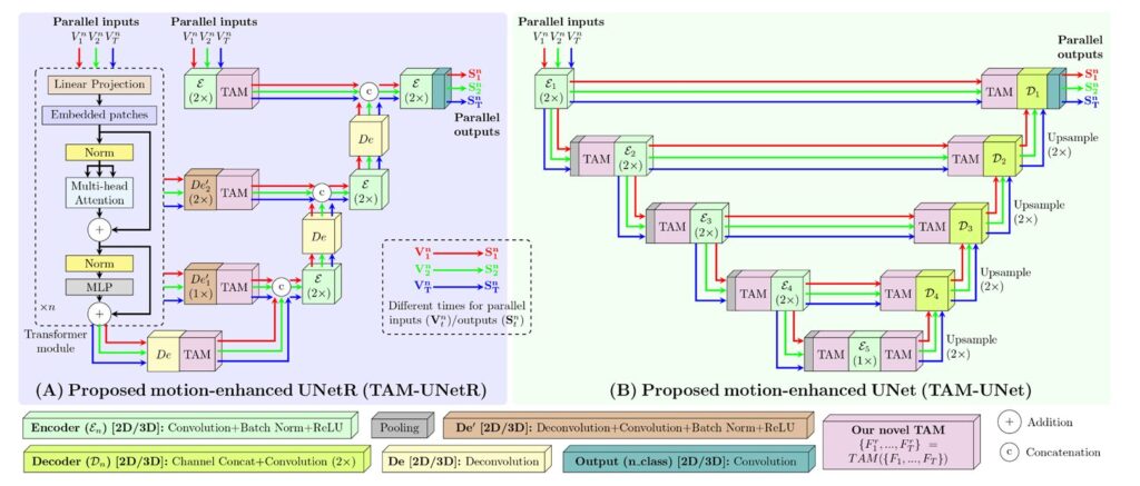
TAM: Plug-and-Play Temporal Attention Module for Motion-Guided Cardiac Segmentation | MedAI Research MedAI Research machine Learning About Cardiac AI · Medical Image Analysis, 2026 · 17 min read The Plug-and-Play Module That Taught Neural Networks to Watch the Heart Move A compact temporal attention module called TAM quietly outperforms much heavier architectures on cardiac segmentation […]
TAM: Plug-and-Play Temporal Attention Module for Motion-Guided Cardiac Segmentation Read More »