AI in medical imaging

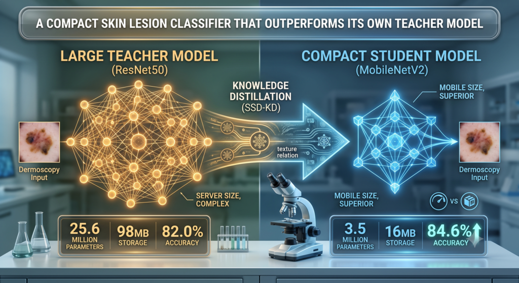
SSD-KD: A Compact Skin Lesion Classifier That Outperforms Its Own Teacher Model

SSD-KD: A Compact Skin Lesion Classifier That Outperforms Its Own Teacher Model

Skin cancer detection has become one of the clearer success stories for deep learning in medicine, with models trained on large dermoscopy image collections repeatedly matching or approaching dermatologist level accuracy on benchmark datasets. The catch is that the models…

SSD-KD: A Compact Skin Lesion Classifier That Outperforms Its Own Teacher Model Read More »

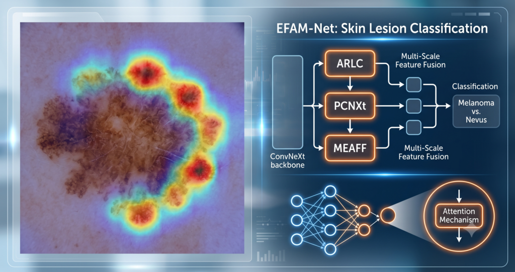
7 Revolutionary Breakthroughs in Skin Cancer Detection: How a New AI Model Outperforms Experts (And Why Older Methods Fail)

7 Revolutionary Breakthroughs in Skin Cancer Detection: How a New AI Model Outperforms Experts (And Why Older Methods Fail)

Skin cancer is one of the most common—and most deadly—forms of cancer worldwide. If detected at an advanced stage, melanoma, the most fatal type, has a 10-year survival rate of less than 39%. But here’s the hopeful news: early detection…

7 Revolutionary Breakthroughs in Skin Cancer Detection: How a New AI Model Outperforms Experts (And Why Older Methods Fail) Read More »

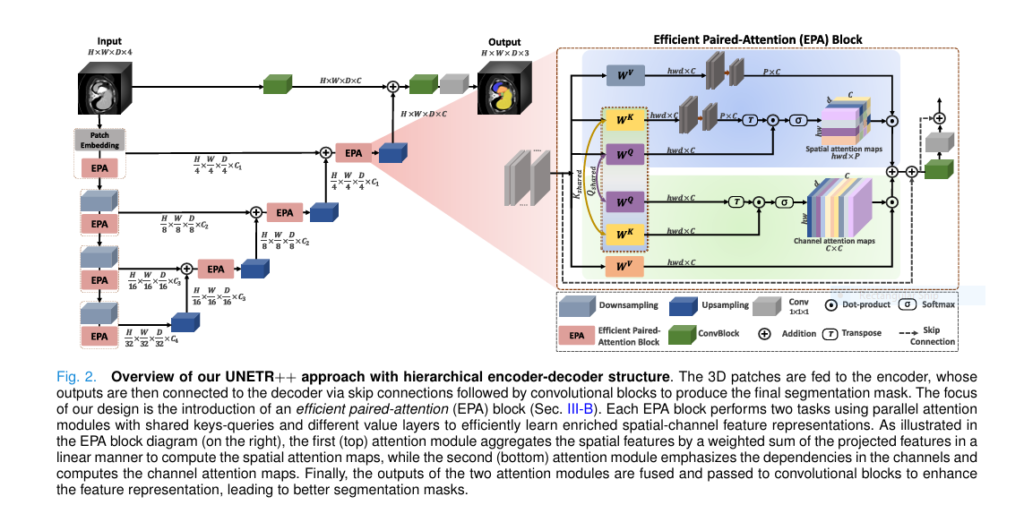
UNETR++ outperforms traditional 3D medical image segmentation methods with 71% fewer parameters and higher accuracy.

UNETR++ vs. Traditional Methods: A 3D Medical Image Segmentation Breakthrough with 71% Efficiency Boost

Medical imaging has always been a cornerstone of diagnostics, treatment planning, and disease monitoring. Among the most critical tasks in this field is 3D medical image segmentation , which enables precise delineation of anatomical structures and pathological regions in volumetric…

UNETR++ vs. Traditional Methods: A 3D Medical Image Segmentation Breakthrough with 71% Efficiency Boost Read More »

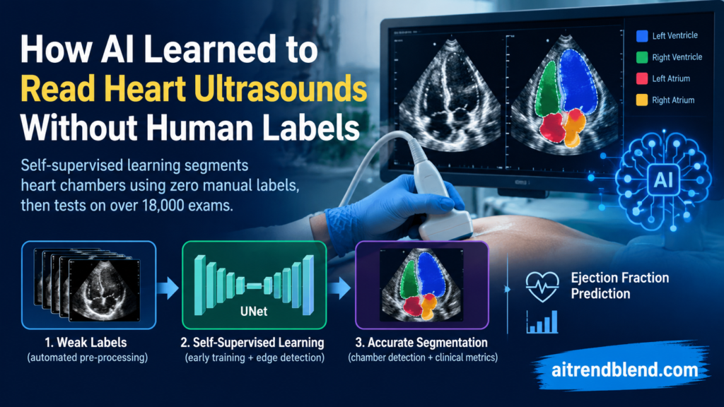
Self Supervised Learning: How AI Learned to Read Heart Ultrasounds Without Human Labels

Self Supervised Learning: How AI Learned to Read Heart Ultrasounds Without Human Labels

Every echocardiogram report that lists an ejection fraction, a chamber volume, or a wall thickness represents a human being drawing a line around a moving, low contrast, occasionally noisy structure on ultrasound. Clinical guidelines from the American Society of Echocardiography…

Self Supervised Learning: How AI Learned to Read Heart Ultrasounds Without Human Labels Read More »