Towards Trustworthy Breast Tumor Segmentation in Ultrasound Using AI Uncertainty
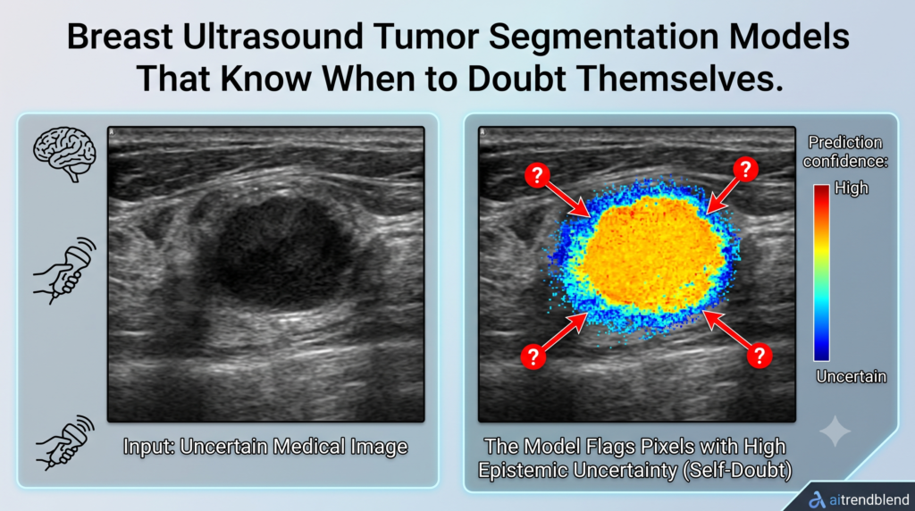
Analysis by the aitrendblend editorial team · Source paper arXiv:2508.17768 Medical Imaging Segmentation Uncertainty Estimation Breast Ultrasound nnU-Net An ultrasound frame next to the kind of entropy map the model produces when it is asked to also grade its own confidence. A radiologist scanning a breast for a suspicious mass rarely gets a clean answer […]
Towards Trustworthy Breast Tumor Segmentation in Ultrasound Using AI Uncertainty Read More »