AI in medical imaging

Diagram illustrating the DIOR-ViT architecture for differential ordinal classification in pathology images

7 Astonishing Ways DIOR-ViT Transforms Cancer Grading (Avoiding Common Pitfalls)

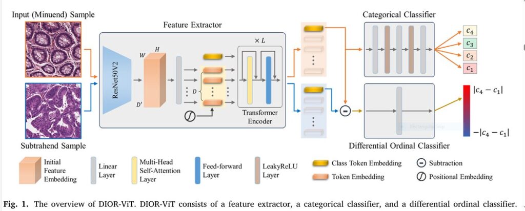
Cancer grading in pathology images is both an art and a science—and it’s riddled with subjectivity, inter-observer variability, and technical roadblocks. Enter DIOR-ViT, a groundbreaking differential ordinal learning Vision Transformer that shatters conventions and delivers robust, high-accuracy cancer classification across multiple tissue types. In this deep-dive SEO-optimized guide, we unpack the seven game-changing innovations behind […]

7 Astonishing Ways DIOR-ViT Transforms Cancer Grading (Avoiding Common Pitfalls) Read More »

Hierarchical Vision Transformers (H-ViT) enhancing prostate cancer grading accuracy through AI-driven pathology analysis

7 Revolutionary Insights from Hierarchical Vision Transformers in Prostate Biopsy Grading (And Why They Matter)

Introduction: Bridging the Gap Between AI and Precision Pathology In the evolving landscape of medical imaging, Hierarchical Vision Transformers (H-ViT) are emerging as a game-changer in prostate biopsy grading , offering unprecedented accuracy and generalizability. Traditional deep learning models have struggled with real-world variability, but H-ViTs are setting new benchmarks by combining self-supervised pretraining, weakly

7 Revolutionary Insights from Hierarchical Vision Transformers in Prostate Biopsy Grading (And Why They Matter) Read More »

Illustration of SPCB-Net architecture showing SK feature pyramid, SAP attention module, and bilinear-trilinear pooling layers for skin cancer detection"

7 Revolutionary Advancements in Skin Cancer Detection (With a Powerful New AI Tool That Outperforms Existing Models)

Introduction: A Critical Need for Advanced Skin Cancer Detection Skin cancer is one of the most common and deadly forms of cancer worldwide. According to the Skin Cancer Foundation , 1 in 5 Americans will develop skin cancer in their lifetime , and melanoma alone accounts for more deaths than all other skin cancers combined

7 Revolutionary Advancements in Skin Cancer Detection (With a Powerful New AI Tool That Outperforms Existing Models) Read More »

Diagram showing intra-class patch swap between two images of the same category, illustrating the self-distillation process without a teacher model.

7 Shocking Wins and Pitfalls of Self-Distillation Without Teachers (And How to Master It!)

Introduction In the world of deep learning, especially in computer vision, knowledge distillation (KD) has been a go-to method to compress large models and improve performance. But the classic approach heavily relies on teacher-student architectures, which come with high memory, computational costs, and training complexity. The new research paper “Intra-class Patch Swap for Self-Distillation” proposes

7 Shocking Wins and Pitfalls of Self-Distillation Without Teachers (And How to Master It!) Read More »

Super-resolution ultrasound with multi-frame deconvolution improving microbubble localization

🚀 7 Game-Changing Wins & Pitfalls of Multi-Frame Deconvolution in Super-Resolution Ultrasound (SRUS)

Introduction: A New Era in Ultrasound Imaging Super-resolution ultrasound (SRUS), or Ultrasound Localization Microscopy (ULM), has redefined the boundaries of medical imaging by enabling visualization of microvasculature at a scale previously thought unattainable. Traditional ultrasound methods are limited by diffraction, but SRUS pushes through this barrier by tracking microbubble (MB) contrast agents in vivo. However,

🚀 7 Game-Changing Wins & Pitfalls of Multi-Frame Deconvolution in Super-Resolution Ultrasound (SRUS) Read More »

Disentangled generative model showcasing independent factors of age, ethnicity, and camera in synthetic retinal images

🔍 7 Breakthrough Insights: How Disentangled Generative Models Fix Biases in Retinal Imaging (and Where They Fail)

Introduction: Why Bias in Retinal Imaging Matters More Than Ever Retinal fundus images are crucial in diagnosing conditions from diabetic retinopathy to cardiovascular diseases. But here’s the problem: most AI models trained on retinal images learn the wrong things. Imagine this: a deep learning system that diagnoses ethnicity instead of actual disease features—because the camera

🔍 7 Breakthrough Insights: How Disentangled Generative Models Fix Biases in Retinal Imaging (and Where They Fail) Read More »

Medical AI transforming tumor segmentation with EGTA-KD technology

Revolutionary AI Breakthrough: Non-Contrast Tumor Segmentation Saves Lives & Avoids Deadly Risks

Imagine detecting deadly tumors without injecting risky contrast agents. A revolutionary AI framework called EGTA-KD is making this possible, achieving near-perfect segmentation (90.8% accuracy) on non-contrast scans while eliminating allergic reactions and kidney damage linked to traditional methods. This isn’t futuristic hype – it’s validated across brain, liver, and kidney tumors in major clinical datasets. The Deadly Cost of Current

Revolutionary AI Breakthrough: Non-Contrast Tumor Segmentation Saves Lives & Avoids Deadly Risks Read More »

Counterfactual contrastive learning closes the performance gap between majority and minority scanners, especially where it matters most: in low-data settings and real-world deployments

Title: 5 Powerful Reasons Why Counterfactual Contrastive Learning Beats Traditional Medical Imaging Techniques (And How It Can Transform Your Practice)

Introduction: The Future of Medical Imaging Starts Here Medical imaging has long been a cornerstone of diagnostics, but traditional methods often fall short when it comes to adapting to real-world variability. Enter counterfactual contrastive learning , an innovative framework that’s changing the game by leveraging causal image synthesis to improve model robustness and downstream performance.

Title: 5 Powerful Reasons Why Counterfactual Contrastive Learning Beats Traditional Medical Imaging Techniques (And How It Can Transform Your Practice) Read More »

SVIS-RULEX SFMOV heatmap overlay on a chest X-ray: Red/Orange areas highlight regions of high statistical significance (e.g., mean intensity, skewness, entropy) corresponding to COVID-19 lung opacities, validated by radiologists. Blue areas show less relevant tissue

3 Breakthroughs & 1 Warning: How Explainable AI SVIS-RULEX is Revolutionizing Medical Imaging (Finally!)

For years, artificial intelligence (AI) has promised to revolutionize medical diagnosis, particularly in analyzing complex medical images like X-rays, MRIs, and ultrasounds. Deep learning models consistently achieve superhuman accuracy in spotting tumors, infections, and subtle pathologies. Yet, a critical roadblock remains: the “black box” problem. How does the AI really make its decision? Without transparency, doctors hesitate to

3 Breakthroughs & 1 Warning: How Explainable AI SVIS-RULEX is Revolutionizing Medical Imaging (Finally!) Read More »

ElastoNet: A revolutionary neural network approach to MR Elastography inversion with uncertainty quantification.

ElastoNet 1: The Revolutionary Neural Network for MRE Wave Inversion with Uncertainty Quantification (Pros & Cons)

Introduction: Why ElastoNet Is Changing the Game in Medical Imaging Medical imaging has seen a rapid evolution over the past decade, especially in non-invasive diagnostics. Among these advancements, Magnetic Resonance Elastography (MRE) has emerged as a powerful technique for evaluating tissue stiffness — a key biomarker in diagnosing diseases like liver fibrosis and cancer. However,

ElastoNet 1: The Revolutionary Neural Network for MRE Wave Inversion with Uncertainty Quantification (Pros & Cons) Read More »

Follow by Email
Tiktok