
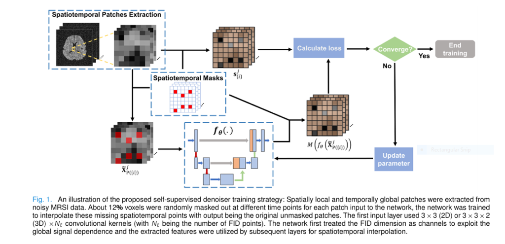
Subspace and Self Supervised Denoising for MRSI Brain Scans
Inside a hospital neuroimaging suite, a patient recovering from a traumatic brain injury who has developed post-traumatic epilepsy (PTE) lies inside a 3 Tesla magnetic resonance scanner equipped with a 20-channel head coil[cite: 49]. Standard structural T1-weighted scans can reveal…
Subspace and Self Supervised Denoising for MRSI Brain Scans Read More »