
Key points
- The method, called DMGSA, trains an unsupervised branch and a supervised branch on the same encoder at the same time rather than pretraining first and fine tuning later.
- A dynamic mask ratio starts easy and grows harder across training, following a sine shaped curve that the authors tie to how a child’s learning difficulty ramps up over time.
- A generalized mean pooling module folds global information about what a foreground airway voxel looks like back into every local feature map before the final prediction.
- Two lightweight discriminators push the network to reconstruct sharper, more realistic textures for the smallest bronchioles, the branches most prone to going missing.
- Trained only on a public dataset of otherwise unremarkable lungs, the network was then tested cold on COVID-19 and lung fibrosis scans, and it still beat every baseline it was compared against on the combined continuity and completeness score.
- The authors are candid that some of the remaining false positives trace back to incomplete ground truth labels rather than to genuine model errors.
Why airway segmentation keeps breaking
Pulmonary fibrosis causes about one percent of all deaths in the United Kingdom, and the paper opens by pointing at a specific gap in how those patients get monitored. Clinicians have no reliable way to predict, from a first scan, whether a given patient’s fibrosis will progress quickly or slowly, which means treatment often waits until the disease has already advanced far enough to see on a follow up scan. High resolution CT is the standard first look, and researchers have spent something like three decades mining it for biomarkers, with traction bronchiectasis severity emerging as one of the more dependable signals across several kinds of fibrotic lung disease. The catch is that visual grading of bronchiectasis is subjective, it varies between readers, and it is not sensitive enough to catch small changes between two scans taken a few months apart.
That is the practical case for an automated airway segmentation tool, and the paper is candid about why building one is hard. Airway voxel intensities are not consistent across scanners and protocols. A single CT volume can contain hundreds of slices where the vast majority of voxels are background, so a naive model quickly learns to ignore the airway altogether because ignoring it barely hurts the loss. Some tracheal walls in the smallest branches are only one or two voxels wide, so a tiny error swallows an entire piece of anatomy. The paper groups the resulting failure modes into three buckets, discontinuity where a traced branch snaps in the middle, false negatives where whole side branches vanish, and leakage where the prediction bleeds outside the true airway wall into surrounding lung tissue.
Prior work attacked pieces of this problem one at a time. Garcia Uceda Juarez and colleagues used a weighted cross entropy loss to rebalance foreground and background voxels. Qin and colleagues, along with a separate effort from Garcia Uceda and coauthors, cropped full CT volumes down to a bounding box around a pre segmented lung to strip out irrelevant background before training. Zheng and colleagues, the team behind a method called WingNet, trained on sampled patches and specifically hunted for hard regions using false negative predictions during training. Wang and colleagues built a bronchiole sensitive loss meant to preserve airway topology across generations of branching, training the network iteratively. On the leakage and discontinuity side, Nadeem and colleagues used a freeze and grow propagation strategy inside a 3D U-Net, Tang and colleagues added a patch scale adversarial refinement step on top of an initial segmentation, and Nan and colleagues, whose FANN method becomes the main point of comparison in this paper, introduced fuzzy attention specifically to fight discontinuity.
Each of those ideas helped, and the paper does not pretend otherwise. What it argues is that none of them fixed the underlying data problem, which is that large scale 3D airway datasets with complete, voxel accurate labels basically do not exist, because the annotation itself takes a trained expert a long time per case and is still imperfect when finished.
Borrowing from masked image modeling, then rebuilding it for 3D lungs
The obvious fix for label scarcity is unsupervised pretraining, and the paper surveys the two broad families that have been tried in medical imaging. Generative approaches, following work like the variational autoencoder line from Luhman and Luhman built on Kingma and Welling’s original formulation, reconstruct images and lean on global redundancy across the volume. Discriminative approaches, inspired by momentum contrast from He and colleagues, try to learn features that are robust and non redundant, but the authors point out a specific mismatch for medical images. Most scans of the same organ look broadly similar to each other, so a contrastive objective built around telling different images apart has less to work with than it does on, say, a dataset of varied natural photographs.
That is what makes masked image modeling attractive instead. He and colleagues’ masked autoencoder, usually shortened to MAE, hides a large fraction of an input image and trains a network to reconstruct the missing pieces, and Wei and colleagues extended the idea to masked feature prediction. But the paper flags two specific problems with applying MAE style pretraining directly to airway CT. First, MAE and similar methods fix the mask ratio at a single high value for the whole run, commonly seventy five or ninety percent, and the authors argue this ignores how humans actually learn, starting with easier tasks and working up to harder ones as skill grows. Second, and more specific to this application, uniform masking treats every voxel as equally informative, when in an airway CT volume the background voxels vastly outnumber the airway voxels and contribute very little signal about bronchiole texture.
There is also an architectural complaint. The dominant MAE recipe follows what the paper calls a pretrain, discard, finetune pipeline shown in the paper’s Figure 3a. You pretrain an encoder and decoder together to reconstruct masked patches, then you throw away the decoder, bolt on a new one suited to the downstream task, and finetune the whole thing on labeled data. The authors call this inefficient for two reasons that matter a lot for a small, expensive to annotate field like airway CT. Discarding a well trained decoder wastes the work that went into training it. And feeding the finetuning stage the full, unmasked image rather than the same masked input the encoder learned on invites overfitting, particularly on datasets this small.
The core design decision
Rather than run unsupervised pretraining first and supervised finetuning second, DMGSA runs both branches on the same encoder and decoder at the same time, from the very first training step, and it never discards or redesigns any part of the network between stages.
How DMGSA is actually put together
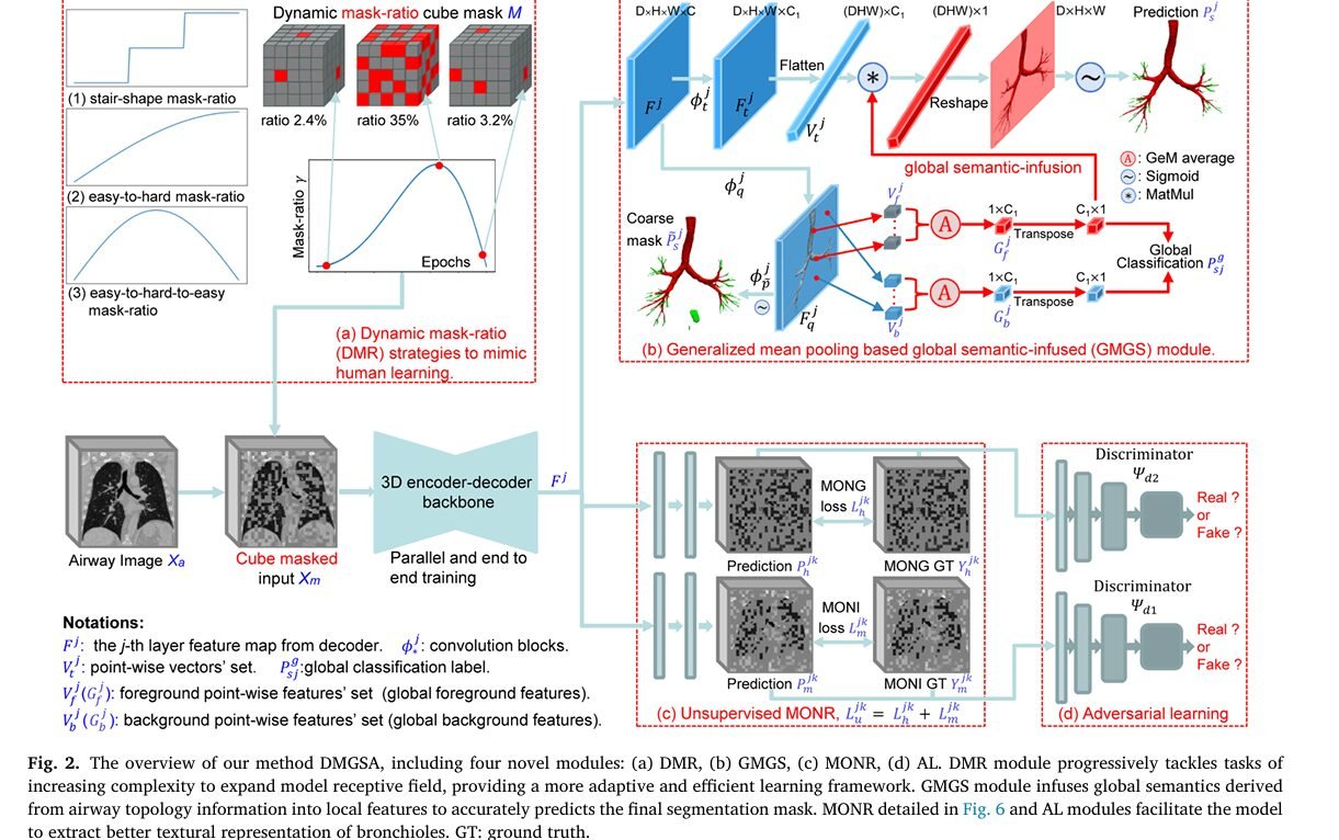
The overall pipeline, shown in the paper’s Figure 2, has five parts working over a 3D CT volume of size depth by height by width. A dynamic mask ratio module first produces a cube masked version of the raw input. A 3D U-shaped encoder decoder backbone, described in the paper’s supplementary material, extracts multi scale hierarchical features from that masked input. Those features then split into two parallel paths. A supervised branch, strengthened by the new generalized mean pooling module, predicts the final airway segmentation. An unsupervised branch predicts a pair of texture targets the authors call multi order normalized responses. Finally, two small discriminator networks compare the predicted texture maps against their ground truth counterparts in an adversarial setup. Every one of these pieces trains together, end to end, in one pass.
Masking that gets harder as training goes on
Instead of the fixed high mask ratio used in classic MAE, the authors test three dynamic schedules shown in their Figure 2a, a stair shape ratio that jumps in discrete steps, an easy to hard ratio that climbs and stays high, and an easy to hard to easy ratio that climbs and then falls back down near the end of training. After running the comparison, the third option won, and it is defined by a fairly compact formula.
The intuition is that a sine curve raised to a power and scaled by pi naturally rises from zero, peaks, and falls back toward zero, so the network sees almost no masking at the very start and the very end of training, with the hardest, most heavily masked examples clustered in the middle. Ablation testing pinned the best power at rho equals two point zero and the best ceiling at r equals zero point three five, which the authors note is a much smaller maximum mask ratio than the seventy five or ninety percent typically used for MAE style pretraining on natural images. Their explanation is that a dense, pixel level segmentation task needs more visible context to learn from than a coarse pretraining objective does. There is a second, more practical payoff to letting the mask ratio fall back toward zero near the end of training. At test time the model sees the full, unmasked scan with no masking at all, so shrinking the mask ratio during late training quietly closes the gap between what the network trained on and what it will actually see in deployment.
Folding global context back into local predictions
The generalized mean pooling based global semantic infused module, shortened to GMGS in the paper, tries to solve the class imbalance problem a different way, by making sure the network never loses sight of what an airway voxel is supposed to look like in aggregate, even while it is busy predicting one voxel at a time. For each decoder layer, two separate convolution blocks produce two feature maps. One gets flattened into point wise vectors. The other gets split, using the ground truth mask, into a set of foreground vectors and a set of background vectors. Those two sets then pass through a trainable generalized mean pooling operation.
The geometric argument for why this helps is almost visual. Two feature vectors both describing airway foreground point in a similar direction in feature space, so their dot product stays high. A foreground vector and a background vector point in fairly different directions, so their dot product drops much further, because cosine similarity falls off quickly as the angle between two vectors widens. By pooling the foreground and background vectors separately into global summaries and then multiplying those summaries back against the local per voxel features, the network sharpens the contrast between what looks like airway and what does not, before the final sigmoid turns that into a probability. The design also adds two extra supervision signals during training, a global classification label and a coarse segmentation mask, both compared against ground truth, which the ablation results below show carries real weight.
Multi order texture targets for the unsupervised branch
The third piece borrows an idea from a recent image retrieval method called SuperGlobal, which found that raising an image to different exponential powers highlights different frequency content and sharpens contrast. The authors apply the same trick to raw airway CT and to gradient maps computed from it, producing what they call multi order normalized images and multi order normalized orientation gradients, together shortened to MONR. The raw volume first gets rescaled to the zero to two hundred fifty five range, then raised to a power k drawn from zero point five or one point five, then renormalized.
Because computing true 3D orientation gradients directly is difficult, the authors instead apply 2D Sobel kernels separately along the depth, width and height axes of each of these reweighted volumes and concatenate the results, producing gradient maps that emphasize the high frequency texture around thin bronchiole walls while suppressing the flatter, more redundant background. Two design choices here are worth calling out because they mark a real departure from standard MAE. Only one, single order version of the input is masked and fed to the encoder, but the network has to predict multiple differently ordered targets from that single masked input in parallel, rather than the usual single input to single output pretraining recipe. And the reconstruction loss is only computed inside the masked regions, which the authors say preserves variety in what gets reconstructed and helps guard against overfitting.
Two small discriminators for extra texture pressure
On top of the MONR targets, the paper adds a lightweight adversarial learning step. Two discriminator networks, one for the image side of MONR and one for the gradient side, each built from five blocks of convolution, instance normalization, and leaky ReLU with a dropout rate of zero point three, learn to tell a real ground truth texture map from a predicted one. The segmentation and reconstruction network, in turn, is trained to fool those discriminators. The two halves optimize a standard minimax objective, with the discriminators trying to maximize their own classification loss on real versus fake patches while the main network tries to minimize it, weighted by balance parameters the authors set to zero point one each. The stated goal is narrow and specific, pushing the network to spend extra effort getting the texture of the smallest, terminal bronchioles right, rather than settling for a blurry but low loss average reconstruction.
What the supervised branch predicts, and how it is scored
Unlike the unsupervised branch, which gets dropped entirely at test time, the supervised branch is what actually ships the final segmentation. During training it predicts the full airway mask directly from the cube masked input at four different decoder depths, and each of those four predictions gets its own combined Dice and binary cross entropy loss, weighted by balance parameters that grow across the layers from zero point one up to zero point five, putting the most weight on the final, highest resolution prediction. At test time the mask disappears and the network simply segments the full, unmasked scan in one pass, with no separate finetuning stage required.
Data, metrics, and how the experiment was set up
Training and validation used the public BAS dataset, ninety cases split fifty four for training, eighteen for validation, and eighteen for testing, built from twenty cases in the EXACT’09 challenge dataset and seventy labeled cases from the LIDC dataset. Two additional datasets were reserved purely for out of distribution testing and never touched during training, twenty five HRCT scans from COVID-19 patients admitted to Wuhan Renmin Hospital, and twenty five lung fibrosis cases drawn from the Open Source Imaging Consortium, with expert labels supplied by specialists at the Royal Brompton Hospital. That is a deliberate stress test, training on relatively typical lungs and then asking the network to hold up on two of the more visually distorted disease patterns it is meant to help with clinically.
Volumes were cropped to one hundred twenty eight by ninety six by one hundred forty four voxel patches during training, chosen so each patch kept at least ten percent airway foreground, with a batch size of four across four NVIDIA V100 GPUs for one hundred twenty epochs. Most of the network trained with AdamW, an initial learning rate of one times ten to the minus three, a weight decay of five times ten to the minus four, gradient clipping, and a cosine learning rate schedule, while the two discriminators used a smaller initial learning rate of one times ten to the minus four. The adversarial balance parameters alpha and beta from the minimax loss were both set to zero point one.
Performance was judged on five metrics that, taken together, are meant to catch the specific failure modes the introduction complains about. Intersection over union and precision measure raw overlap with the ground truth mask and tend to be dominated by the trachea and the thick central bronchi simply because those regions have far more volume. Detected branch ratio and detected length ratio instead measure what fraction of individual branches, and what fraction of total branch length, were correctly recovered, which is where thin, easily missed bronchioles actually show up in the score. Airway missing ratio tracks the volume of false negatives relative to the ground truth. And a combined continuity and completeness F score, abbreviated CCFs, blends intersection over union with detected length ratio using a weighting term the authors set to zero point nine, specifically so a model cannot post a high score just by nailing the big trachea while quietly losing the branch tips.
The headline numbers
On the BAS test set DMGSA posted an intersection over union of eighty eight point zero two percent, precision of ninety two point one five percent, detected length ratio of ninety six point zero four percent, detected branch ratio of ninety five point one one percent, airway missing ratio of five point six one percent, and a CCFs of ninety one point three five percent, which was the best CCFs, detected length ratio, and detected branch ratio among every method compared, edging out FANN’s combined score of eighty nine point six nine percent by one point six six points. It is worth being precise about what it did not win. NaviAirway posted a lower airway missing ratio at four point one three percent, and V-Net edged it out on raw precision at ninety seven point eight one percent, but both of those methods trailed badly on detected branch ratio, at eighty one point zero one percent and twenty two point zero four percent respectively, which is exactly the kind of tradeoff the CCFs metric was built to expose.
The COVID-19 dataset, never seen during training, is where the generalization argument gets tested directly. DMGSA reached ninety three point three one percent intersection over union, ninety seven point four nine percent detected length ratio, ninety seven point zero one percent detected branch ratio, and a CCFs of ninety five point one three percent, a two point four three point gain in CCFs over FANN’s ninety two point seven percent, with a smaller standard deviation too, which the authors read as a sign of steadier performance across the twenty five cases rather than just a higher average pulled up by a few easy scans.
The lung fibrosis dataset is described in the paper as the hardest of the three, and the gap widens further there. DMGSA reached eighty four point zero one percent intersection over union, eighty seven point one two percent detected length ratio, eighty two point one seven percent detected branch ratio, six point zero three percent airway missing ratio, and a CCFs of eighty five point two eight percent, a four point two nine point improvement in CCFs over FANN’s eighty point nine nine percent. For comparison, the older V-Net baseline managed only a CCFs of fifteen point seven six percent on this dataset, a reminder of just how badly fibrotic distortion breaks methods that were not built with thin branch continuity specifically in mind.
| Dataset | FANN CCFs | DMGSA CCFs | Gain |
|---|---|---|---|
| BAS test set | 89.69% | 91.35% | +1.66 points |
| COVID-19 (unseen during training) | 92.70% | 95.13% | +2.43 points |
| Lung fibrosis (unseen during training) | 80.99% | 85.28% | +4.29 points |
What the ablation studies actually show
The most convincing part of the paper, at least for a reader trying to judge whether each new module earns its place, is the module by module ablation run on the lung fibrosis dataset in the paper’s Table 6. Starting from a reproduced FANN baseline with a CCFs of eighty one point one three percent, adding the direct masked input strategy alone lifted CCFs by zero point six seven points and, notably, produced the single highest precision of any row at eighty nine point one five percent. Layering in the dynamic mask ratio on top of that produced the largest individual jump in the whole table, one point one two CCFs points, which lines up with the authors’ argument that gradually widening the receptive field genuinely helps the network learn long range branch topology rather than just memorizing local patterns.
Adding the GMGS module next contributed a zero point nine five point CCFs gain while cutting the airway missing ratio by the largest single margin in the table, zero point four seven points, consistent with the module’s stated job of pushing global foreground and background context back into every local prediction. The MONR unsupervised targets added another one point zero two CCFs points, and the adversarial learning step contributed a smaller zero point three nine points on top of that, though the authors note it still produced a visible improvement specifically in detected length ratio and detected branch ratio, meaning it was doing exactly the narrow job it was designed for even though its overall score contribution looks modest next to the other three modules.
A second ablation, comparing this parallel training scheme against a more conventional serialized pretrain then finetune pipeline built as a fair, same architecture MAE style baseline, is arguably the paper’s strongest evidence for its central design claim. That serialized baseline, referred to as MAE with a dagger in Table 8, needed one hundred fifty training epochs and produced a zero point eight four CCFs point gain over the same starting baseline. The direct masked input approach alone reached a comparable region in fewer epochs, one hundred twenty, and once the parallel single order training scheme was added on top, the combined approach reached a zero point nine six CCFs point gain, edging out the serialized baseline while training in a single stage instead of three separate ones. That is a real, if modest, efficiency and performance argument for skipping the pretrain, discard, finetune pattern altogether in a data scarce field like this one.
A third ablation drilled into the MONR module’s two sub targets separately. Adding only the multi order normalized image target produced a zero point four four CCFs gain but a much larger jump in detected length ratio, one point zero six points, and detected branch ratio, one point seven five points, suggesting it specifically helps with discontinuity and missed branches rather than raw overlap. Adding only the gradient based multi order normalized orientation gradient target instead produced a larger one point zero two CCFs gain along with bigger detected length ratio and detected branch ratio jumps, two point one seven and three point three one points respectively, and the largest airway missing ratio reduction of the two, zero point five two points. Using both targets together, as the full method does, reached a combined one point two one CCFs gain, more than either target managed alone.
Smaller sweeps rounded out the picture. Testing different exponent orders for the MONR targets confirmed that using all three orders together, zero point five, one point zero, and one point five, beat any single order or narrower combination. Testing all three dynamic mask ratio schedules against a fixed static ratio confirmed that every dynamic option beat the static baseline, with the easy to hard to easy schedule winning outright, which the authors attribute specifically to that schedule shrinking the mask ratio back toward zero near the end of training and thereby narrowing the gap between the masked training distribution and the fully visible test distribution. A separate sweep over the maximum mask ratio r found zero point three five to be the sweet spot, and a sweep over the shape parameter rho in the sine schedule found two point zero to work best.
Reading the qualitative results honestly
The paper includes rendered 3D airway trees for two cases each from BAS, COVID-19, and lung fibrosis, comparing SFCN, WingNet, FANN, and DMGSA side by side, with true positive voxels in red, false positives in green, and false negatives in blue. The pattern the authors describe holds up across the figure. SFCN shows large, obvious green blocks of leaked prediction bleeding outside the true airway wall. WingNet tends to lose the main trachea itself in blue, which drags its overall overlap score down even though it can pick out some fine branches elsewhere. FANN generally tracks the branching structure well but shows small blue gaps of discontinuity in the terminal bronchioles, and on the harder lung fibrosis cases it picks up a more serious green leakage problem. DMGSA’s renderings show visibly fewer blue gaps and less green leakage across all three datasets, though the authors are transparent that it is not spotless.
A limitation the authors themselves flag
Some of the remaining false positives in DMGSA’s predictions trace back not to genuine model mistakes but to a minority of imperfect ground truth annotations, cases where a human annotator missed a terminal bronchiole that the model actually found. The paper illustrates this directly in its Figure 11, where correctly predicted airway voxels sit outside the red ground truth region simply because the label itself is incomplete.
Clinical translation gap
There is a meaningful distance between a strong CCFs score on a curated research dataset and a tool a radiology department could actually rely on day to day. The paper’s own test set for the most clinically relevant conditions is small, twenty five COVID-19 scans and twenty five lung fibrosis scans, each drawn from a single institution’s imaging protocol and a single set of expert annotators at the Royal Brompton Hospital. That is enough to demonstrate that the method generalizes better than several published alternatives, but it is not the scale or the multi site diversity a regulatory submission or a multi center clinical validation study would need. The paper also does not report inference time, memory footprint, or how the segmentation output would integrate into an existing radiology workflow, all of which matter more once a research result becomes a piece of deployed software. And because the ground truth annotations themselves are demonstrably imperfect, as the authors acknowledge with their own example in Figure 11, any clinical deployment would need a validation study measured against a more rigorously adjudicated reference standard, not just against the same imperfect labels the model was scored against here.
Clinical and dataset limitations
Beyond the translation gap, a few specific constraints are worth naming plainly. The training data comes from ninety cases total, split fifty four, eighteen, and eighteen across train, validation, and test, which is a small sample by the standards of most deep learning applications, even though it is a fairly typical size for 3D medical segmentation where annotation cost is high. The two out of distribution test sets, twenty five cases each for COVID-19 and lung fibrosis, are similarly modest, and both were sourced from specific hospitals, Wuhan Renmin Hospital and the Open Source Imaging Consortium respectively, which limits how confidently the reported numbers generalize to scanners, protocols, and patient populations the network has never encountered. The paper does not report results broken out by fibrosis severity or by disease subtype, so it is not possible from the numbers given to say whether performance holds up equally well across the full range of how advanced a patient’s fibrosis might be. Readers should treat the reported metrics as evidence that the architecture generalizes better than the compared baselines on these specific datasets, not as a claim about performance in a broader, unstudied population.
Where this fits in the bigger picture
Zoomed out, the paper’s real argument is less about airway CT specifically and more about how to structure learning when labels are scarce and background noise dwarfs the signal you actually care about, which describes a lot of medical imaging problems beyond the lungs. The parallel training idea, running an unsupervised reconstruction objective and a supervised segmentation objective on one shared backbone at once instead of in two separate stages, is not tied to airway anatomy at all, and the dynamic mask ratio schedule is a fairly general recipe that could plausibly transfer to other sparse, thin structure segmentation tasks such as vasculature, nerve fibers, or other branching anatomical trees. The multi order texture targets lean specifically on the observation that thin, low contrast structures respond differently to different exponential rescalings of the raw intensity, which again is not a lungs only phenomenon.
Limitations and open questions
The authors’ own weakness section is short and specific, focused on the false positive issue tied to incomplete annotations discussed above, but a few broader open questions are worth naming for anyone evaluating this work critically. The comparison baselines were reproduced by the authors themselves in most cases rather than run by the original authors of each method, which is standard practice but always introduces some risk that a baseline was not tuned as carefully as the proposed method. The GeM power index alpha and the mask ratio schedule parameters rho and r were tuned empirically on this specific dataset combination, and it is not yet clear from the paper how sensitive those settings would be if applied to a very differently sized or differently annotated dataset. And while the paper reports standard deviations alongside every mean, which is good practice, it does not report confidence intervals or a full statistical power analysis for the smaller twenty five case test sets, so the precision of the reported gains on those datasets specifically should be read with some caution.
Frequently asked questions
What does DMGSA stand for and what problem does it solve
DMGSA stands for Dynamical Multi order responses and Global Semantic infused Adversarial network. It is a deep learning method for automatically tracing the full tracheobronchial tree on chest CT scans, built specifically to work well even when very few scans have complete, voxel level hand drawn labels.
How is this different from a standard masked autoencoder approach
Classic masked autoencoder methods mask a fixed, usually large, fraction of the input throughout a single pretraining run, then discard the pretraining decoder and finetune a new one on labeled data in a separate stage. DMGSA instead uses a mask ratio that grows and then shrinks across training, and it trains its unsupervised and supervised objectives together on the same encoder and decoder from the first step, with no separate finetuning stage.
Was the model tested on data it had never seen during training
Yes. The network was trained only on the public BAS dataset, built from EXACT’09 and LIDC cases, and then tested without any additional training on twenty five COVID-19 scans and twenty five lung fibrosis scans that it never encountered during training.
Which metric matters most for judging airway segmentation quality
The paper argues that intersection over union alone can be misleading because it is dominated by the large main trachea. It relies more heavily on detected branch ratio, detected length ratio, and a combined continuity and completeness score called CCFs, since those are more sensitive to whether the thinnest terminal bronchioles were traced correctly.
Does this method rely on the discriminator networks used for adversarial learning at test time
No. The two discriminator networks and the entire unsupervised branch that predicts multi order texture targets are only used during training. At test time the network runs only the supervised segmentation branch on the full, unmasked scan.
Is this method ready to be used in a hospital setting
Not based on what is reported in the paper. The clinical test sets are small, drawn from single institutions, and the paper does not report the kind of multi site validation, inference speed, or workflow integration testing that a clinical deployment would require. The authors themselves also note that some of the model’s apparent errors are actually gaps in the human drawn ground truth labels rather than genuine model mistakes.
Read the source paper
The full method, all five ablation tables, and the supplementary architecture details are in the open access paper published in Medical Image Analysis.
Conclusion
The core achievement here is narrower and more useful than it might first sound. DMGSA is not claiming to have solved airway segmentation outright, and the authors do not use that kind of language anywhere in the paper. What it demonstrates instead is that a specific set of training choices, a mask ratio that ramps up and back down instead of staying fixed, a pooling scheme that keeps global foreground and background context in view during local prediction, texture targets computed at multiple exponential orders, and a light adversarial nudge toward sharper reconstructions, add up to a meaningfully better result on the exact metrics built to catch the failure modes that matter clinically, detected branch ratio and detected length ratio for the smallest bronchioles, rather than just raw overlap on the big central trachea.
The conceptual shift worth remembering is the move away from pretrain, discard, finetune as the default recipe for combining unsupervised and supervised learning in a small labeled dataset regime. The ablation comparing a serialized MAE style baseline against the parallel training scheme is not a huge margin, well under one CCFs point, but it is evidence in a direction that runs against a fairly entrenched habit in the field, and it comes with a genuine efficiency argument since the parallel approach needed fewer training epochs to get there.
Whether the specific formulas transfer cleanly to other organs and other thin branching structures is an open question the paper does not answer, since every experiment here stays within chest CT and airway anatomy. But the underlying pattern, extremely sparse foreground, catastrophic sample imbalance, and label scarcity driven by expensive expert annotation, describes a long list of other segmentation problems in medical imaging, from retinal vasculature to peripheral nerve tracing to coronary artery trees, and the dynamic masking and multi order texture ideas do not appear to depend on anything specific to lungs.
The honest limitations are worth restating plainly rather than glossing over. The clinical test sets are small and single institution. The comparison baselines were mostly reproduced rather than independently verified against the original authors’ own results. And the authors themselves point out that a chunk of the model’s apparent remaining errors are actually gaps in imperfect human labels, which is a useful reminder that a segmentation benchmark score is only ever as good as the ground truth it is measured against.
For a field where the annotation bottleneck is the single biggest obstacle to progress, a method that gets meaningfully further on the same small labeled dataset, and does it with one training stage instead of three, is worth paying attention to even before it becomes something a hospital could actually deploy.
Proposed model implementation in PyTorch
The following is an independent, illustrative PyTorch implementation of the core ideas described in the paper, the dynamic mask ratio schedule, a simplified 3D encoder decoder backbone, the GMGS module, the multi order normalized response targets, and the lightweight discriminators, wired together with the loss functions described in Equations 3 through 15 of the paper. It is written to run end to end on small random dummy volumes as a smoke test and is meant for learning the architecture, not for clinical use.
Academic citation. Zhang, S., Nan, Y., Fang, Y., Wang, S., Liu, Y., Papanastasiou, G., Gao, Z., Li, S., Walsh, S., and Yang, G. Dynamical multi order responses and global semantic infused adversarial learning, a robust airway segmentation method. Medical Image Analysis, volume 108, article 103867, 2026. Published open access under a CC BY license.
This analysis is based on the published paper and an independent evaluation of its claims.
Thanks for sharing. I read many of your blog posts, cool, your blog is very good. https://accounts.binance.com/register-person?ref=JW3W4Y3A