Medical AI

Medical AI brings together our reporting on machine learning for clinical and biomedical problems, from diagnosis and prognosis to medical image analysis and decision support. Because mistakes in this domain carry a human cost, we pay close attention to evaluation, calibration, uncertainty, and the gap between benchmark numbers and bedside reliability. Expect grounded explainers of recent research rather than uncritical product announcements.

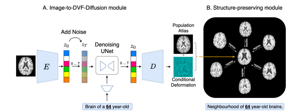
Diff-Def: Diffusion-Generated Deformation Fields for Conditional Brain Atlases.

Diff-Def: Diffusion-Generated Deformation Fields for Conditional Brain Atlases

Researchers from TU Munich and Imperial College London built a latent diffusion model that generates deformation fields — not intensities — to morph a general population brain atlas into condition-specific ones, producing sharper, more anatomically faithful, and fully interpretable atlases…

Diff-Def: Diffusion-Generated Deformation Fields for Conditional Brain Atlases Read More »

Clinical LLMs 2026: Med-Gemini, Med-PaLM 2, and GPT-5 in Medicine.

Clinical LLMs 2026: Med-Gemini, Med-PaLM 2, and GPT-5 in Medicine

Dr. Fatima Nkosi is managing 19 patients on a Saturday night hospitalist shift. At 2am, she opens the clinical AI assistant embedded in her hospital’s Epic instance and types: “64-year-old male, day 2 post-op colectomy, HR 118, temp 38.9°C, WBC…

Clinical LLMs 2026: Med-Gemini, Med-PaLM 2, and GPT-5 in Medicine Read More »

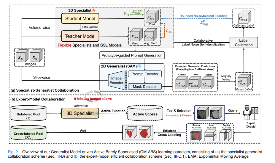
GM-ABS: SAM-Driven Active Barely Supervised 3D Medical Image Segmentation.

GM-ABS: SAM-Driven Active Barely Supervised 3D Medical Image Segmentation

Researchers from CUHK and Harvard Medical School built a training paradigm where the in-training specialist autonomously crafts prompts for a frozen SAM generalist, which returns noisy-but-useful labels across three orthogonal views — while active learning selects the most informative scans…

GM-ABS: SAM-Driven Active Barely Supervised 3D Medical Image Segmentation Read More »

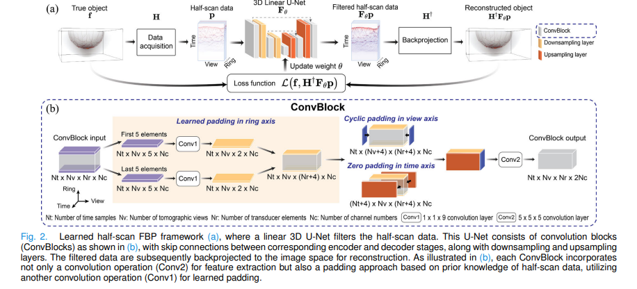
Learned Half-Scan FBP: Physics-Informed Deep Learning for PACT Breast Imaging

University of Illinois researchers solved a decade-old open problem in photoacoustic breast imaging — how to reconstruct from hemispherical data without a closed-form formula — by training a strictly linear 3D U-Net to approximate the unknown filter, achieving iterative reconstruction…

Learned Half-Scan FBP: Physics-Informed Deep Learning for PACT Breast Imaging Read More »

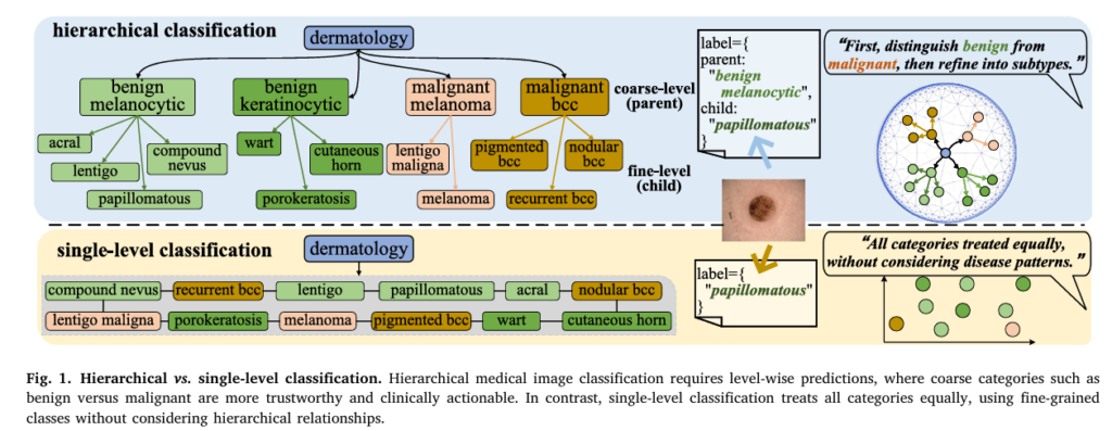
H2CL: Dual-Geometry Hyperbolic-Euclidean Image-Text Learning for Medical Hierarchical Classification.

H2CL: Dual-Geometry Hyperbolic-Euclidean Image-Text Learning for Medical Hierarchical Classification

A UNSW Sydney team built H²CL — a dual-geometry image-text framework that simultaneously operates in Euclidean and hyperbolic spaces, combining group contrastive learning with a hyperbolic entailment loss, to classify medical images across clinical taxonomies. The result: 7% accuracy gains…

H2CL: Dual-Geometry Hyperbolic-Euclidean Image-Text Learning for Medical Hierarchical Classification Read More »

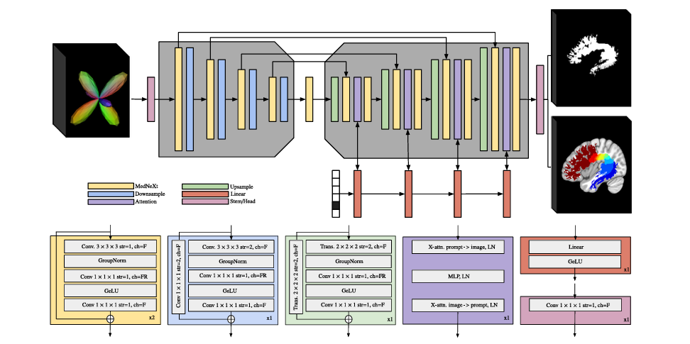
BundleParc: Tractography-Free White Matter Bundle Parcellation with MedNeXt + Cross-Attention

BundleParc: Tractography-Free White Matter Bundle Parcellation with MedNeXt + Cross-Attention

Researchers at Université de Sherbrooke built a prompt-conditioned MedNeXt encoder-decoder that reads fiber orientation maps and outputs anatomically consistent white matter parcellations in seconds — without generating a single streamline — outperforming all competing methods on reproducibility across healthy subjects,…

BundleParc: Tractography-Free White Matter Bundle Parcellation with MedNeXt + Cross-Attention Read More »

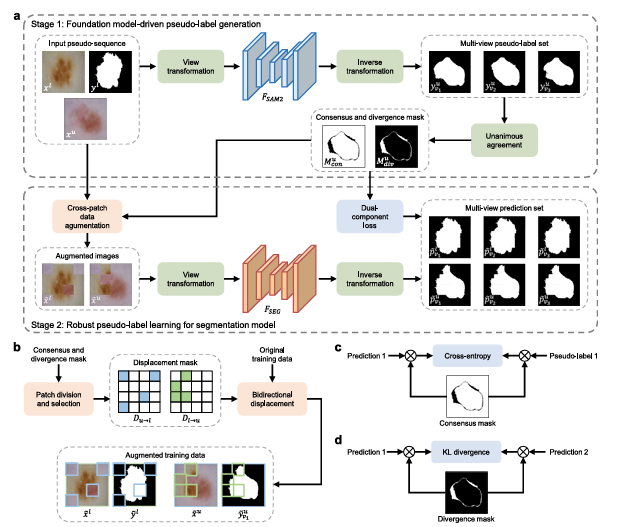
YoloSeg: One Labeled Image Is All You Need for Medical Image Segmentation.

YoloSeg: One Labeled Image Is All You Need for Medical Image Segmentation

A team at the Chinese Academy of Sciences built a framework that achieves segmentation accuracy within 3% of fully-supervised models across ten diverse medical datasets — using exactly one manually annotated image per task. SAM2 does the label propagation; a…

YoloSeg: One Labeled Image Is All You Need for Medical Image Segmentation Read More »

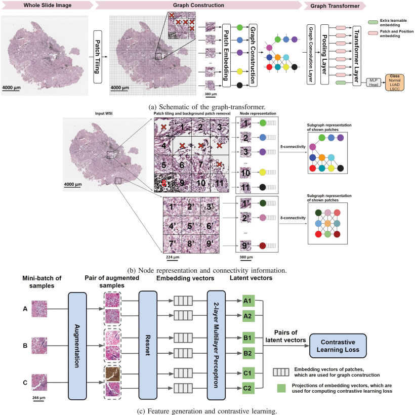
GTP: The Graph-Transformer That Reads Whole Slide Pathology Images Like a Pathologist

GTP: The Graph-Transformer That Reads Whole Slide Pathology Images Like a Pathologist

A team at Boston University built GTP — a Graph-Transformer for Pathology that fuses graph convolutional networks with vision transformers to classify gigapixel whole slide images of lung cancer at 91.2% accuracy, plus a novel GraphCAM technique that highlights exactly…

GTP: The Graph-Transformer That Reads Whole Slide Pathology Images Like a Pathologist Read More »