
Key points
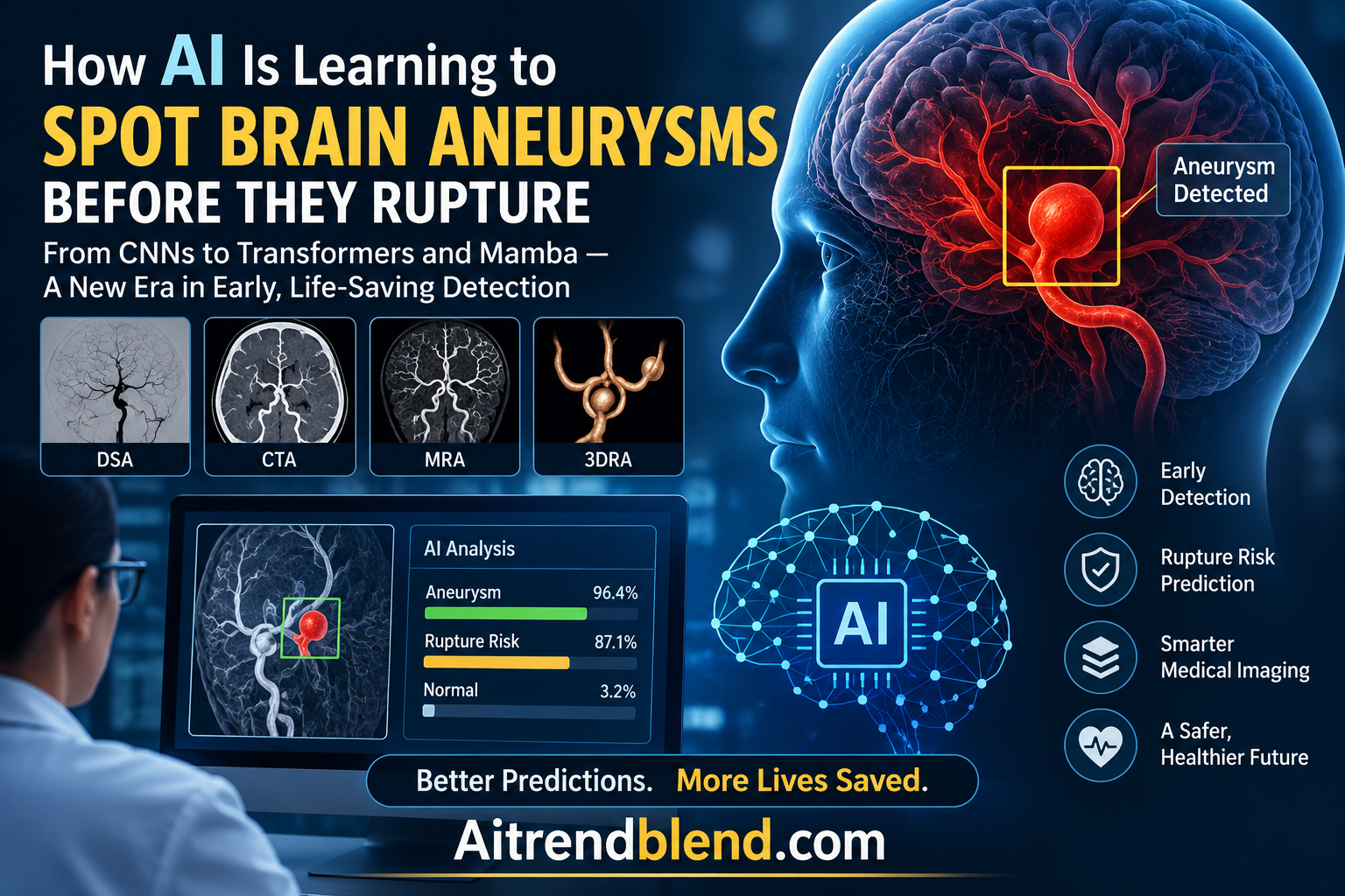
- The survey organizes deep learning research on intracranial aneurysms into three tasks, segmentation, detection, and rupture risk prediction, across four imaging modalities, DSA, 3DRA, CTA, and MRA.
- The field has moved through five architectural eras since 2010, from handcrafted filters to CNNs, then hybrid CNN-Transformer models, geometric deep learning on non-Euclidean vessel surfaces, and now Mamba based state space models and medical foundation models.
- Public datasets remain scarce. The authors catalog eleven public datasets across all modalities plus three synthetic ones, and note that DSA in particular has historically lagged behind CTA and MRA in available benchmarks.
- Rupture risk prediction is shifting from single modality morphology toward multimodal frameworks that combine imaging, radiomics, clinical variables, and computational fluid dynamics.
- Despite strong reported performance, with some models reaching AUC values above 0.96 in individual studies, the authors identify persistent gaps in detecting aneurysms smaller than 3mm and in translating any of this work into real clinical workflows.
Why catching an aneurysm early is so hard
An aneurysm is a weak spot in an artery wall that balloons outward under blood pressure. When one forms near or inside the brain, it usually causes no symptoms at all, which is exactly what makes it dangerous. If it ruptures, the result is a subarachnoid hemorrhage, a medical emergency with a high death rate and long term neurological consequences for many survivors. The size, shape, and location of an aneurysm all factor into how likely it is to rupture, but assessing those factors accurately from a scan is genuinely difficult work.
The imaging itself is only part of the challenge. Aneurysms are small relative to the full scan volume, they often have almost the same brightness as the healthy blood vessels around them, their shapes vary enormously from one patient to the next, and motion artifacts or contrast timing can obscure the exact boundary between a bulge and normal vessel curvature. Put those together and it becomes clear why even experienced neuroradiologists show real variability when reading the same scan, and why there has been such sustained interest in building automated systems that can offer a second, consistent pair of eyes.
Why this matters for AI research broadly
Intracranial aneurysm analysis is a useful stress test for a lot of ideas that show up elsewhere in medical AI, extreme class imbalance, non-Euclidean anatomy that does not fit neatly into a voxel grid, and a genuine shortage of labeled data. How the field has adapted its toolkit here says something about where medical imaging AI is headed more broadly.
Four ways to look inside a blood vessel
The survey covers four main imaging techniques, each with a different balance of invasiveness, resolution, and practical accessibility.
| Modality | Type | Main strength | Main limitation |
|---|---|---|---|
| DSA | Invasive | Gold standard resolution, dynamic flow assessment, catches small or complex aneurysms other scans miss | Visualizes only the injected vessel, radiation and contrast risk, time consuming |
| 3DRA | Invasive | High resolution 3D reconstruction, detailed morphology for wide neck aneurysms | Shares DSA’s invasiveness and radiation exposure, longer procedure time |
| CTA | Noninvasive | Fast, widely available, strong for emergency diagnosis and surgical planning | Ionizing radiation, limited sensitivity for very small aneurysms |
| MRA | Noninvasive | No radiation, good for repeated monitoring and younger patients | Lower spatial resolution, sensitive to motion and flow artifacts |
Digital subtraction angiography, DSA, remains the clinical gold standard, since a contrast agent injected directly into the cerebral vessels followed by digital subtraction of pre and post contrast X-ray images produces extremely clear vascular detail, down to submillimeter precision in identifying micro aneurysms that a 1.5 Tesla MRA scan might miss entirely. But that precision comes at a real cost, DSA is invasive, exposes patients to radiation, and only images the specific artery that was injected, so it is typically reserved for cases where noninvasive screening has already flagged something that needs a closer look. CTA and MRA, by contrast, are the workhorses of initial screening precisely because they do not require an interventional procedure, with CTA favored in emergency settings for its speed and MRA favored for repeated, free of radiation monitoring over time.
A fifth technique, vessel wall imaging, gets its own attention in the survey because it does something the other four cannot. Rather than just showing the shape of the vessel lumen, high resolution vessel wall MRI can visualize the biological state of the aneurysm wall itself, detecting inflammation and enhancement patterns that may signal an unstable, prone to rupture lesion even when its geometry looks identical to a stable one on a standard scan. The catch is that it requires high field MRI equipment, takes considerably longer to acquire, and has far fewer annotated datasets available than the more established modalities.
Where the training data actually comes from
One of the survey’s more sobering contributions is simply cataloging how little public data this field has to work with. The authors identify eleven public clinical datasets spanning DSA, 3DRA, CTA, and MRA, ranging from DIAS, a 2024 DSA dataset with 120 image sequences, to ADAM, a 2020 MRA challenge dataset with 113 training cases. Most current research still relies on private, institutional datasets, which makes cross study comparison difficult and raises real questions about how well any given model would generalize to a hospital it was never trained on.
To help close that gap, three synthetic datasets have emerged recently. Aneumo generates 10,660 synthetic 3D aneurysm shapes from 427 real geometries, each paired with computational fluid dynamics simulations of velocity, pressure, and wall shear stress. AneuG-Flow does something similar at larger scale, offering 14,000 steady flow cases and 200 pulsatile flow cases built from 116 real aneurysm shapes. A third dataset, IAVS, pairs 641 real 3D MRA volumes with synthetic patches and precomputed hemodynamic flow fields. These synthetic sets let researchers control variables like neck width or parent vessel curvature that are hard to sample systematically from real patients, though the authors are careful to flag that synthetic data introduces its own risk of domain shift when a model trained mostly on simulated geometry meets the messier reality of a clinical scan.
Five architectural eras in fifteen years
The survey traces a clear technological arc, visualized in the paper as a timeline running from 2010 to 2026, and it is worth walking through because each stage was a direct response to a limitation of the one before it.
Handcrafted features and early CNNs
Before deep learning took hold, computer aided diagnosis systems relied on rule based algorithms and handcrafted features, intensity thresholding, region growing, active contours, tuned by researchers rather than learned from data. These systems were highly sensitive to variations in scanner protocol and struggled badly with the complex morphology of real vasculature. The U-Net architecture changed that by learning hierarchical features directly from labeled examples, and its encoder decoder structure with skip connections became the default starting point for aneurysm and vessel segmentation, later extended by UNet++ with nested skip connections to better bridge the gap between shallow and deep features.
The shift to attention and Transformers
Pure convolutional networks are good at local pattern recognition but structurally limited in modeling long range spatial relationships, since a convolution only sees a small neighborhood at a time. Hybrid CNN-Transformer models addressed this by combining a convolutional branch for fine grained local detail with a self attention branch that can relate any two points in a scan regardless of distance. Models like SDAN, which introduced a dedicated edge aware attention module and a global shape aware fusion block, showed measurable gains over pure CNN baselines specifically on the vascular overlap and small target problems that plague DSA images. The tradeoff is real, though, since self attention’s computational cost grows quadratically with resolution, and some of the survey’s cataloged models run past 77 million parameters, making them expensive to train and slow to deploy in a time sensitive clinical setting.
Geometric deep learning on the vessel surface itself
A more fundamental rethink came with geometric deep learning. Standard CNNs necessarily process medical images as rigid voxel grids, which introduces discretization error, fine surface detail simply gets lost when a smoothly curving vessel wall gets forced onto a cubic lattice. Geometric deep learning instead works directly on non-Euclidean representations, point clouds, surface meshes, and centerline graphs, preserving the vessel’s actual geometry and topology. The release of the IntrA dataset in 2020, the first large public collection of annotated 3D surface meshes specifically for aneurysms, catalyzed this shift, and subsequent work using architectures like PointNet and PointNet++ demonstrated meaningfully higher accuracy than comparable voxel based baselines for exactly the fine, irregular surface details, like where the aneurysm neck blurs into the parent vessel, that voxelization tends to smear.
Mamba and the return of efficiency
The most recent architectural family in the survey is built on state space models, particularly the Mamba architecture, which can model long range dependencies with linear rather than quadratic computational complexity. This matters enormously for 3D medical volumes, where a Transformer’s quadratic cost becomes genuinely prohibitive at full resolution. MTMU, a Mamba based model built specifically for small aneurysm segmentation, achieves competitive results with just 2.10 million parameters compared to the 13 million parameters of GLIA-Net, a substantial efficiency gain that matters for any hope of running these models close to the point of care rather than on a dedicated research cluster.
From finding the aneurysm to predicting whether it will rupture
Detection and segmentation answer a spatial question, where is the aneurysm and what shape is it. Rupture risk prediction is a fundamentally harder, more clinically consequential question, since most aneurysms never rupture and treating one that would have stayed stable exposes a patient to unnecessary surgical risk, while missing one that will rupture is catastrophic. Traditional clinical scoring systems like PHASES and ELAPSS estimate risk from coarse factors, age, hypertension, lesion size, but the survey traces a clear trend toward richer, learned representations of risk.
Early approaches followed a two stage pipeline, segment the aneurysm and its parent vessel first, then extract morphological features, neck width, dome height, aspect ratio, for a downstream classifier. This structure is interpretable but fragile, since a small segmentation error early in the pipeline can meaningfully distort every risk feature computed afterward. Geometric deep learning models like PMMNet pushed past this by fusing 3D point cloud geometry with multi view 2D representations in a single dual branch network, treating classification and segmentation as a joint rather than sequential problem.
The most recent and, per the survey, most clinically promising direction is multimodal fusion. MCANet combines raw CTA images, radiomics descriptors, morphological measurements, and patient level clinical variables like smoking history and hypertension into a single prediction, and the authors report it maintained strong performance across two independent external patient cohorts despite relatively small sample sizes, a meaningful signal given how often medical AI models fail to generalize outside their original training hospital. A separate framework called IAIT goes further still, fusing CTA and MRA imaging volumes with structured clinical text records through a hierarchical attention mechanism designed specifically to stay sensitive to small lesions that a purely image based model might miss.
Key takeaway
The field is converging on a shared realization, rupture is not purely a geometric event. Two aneurysms that look nearly identical on a scan can behave very differently depending on hemodynamic stress, wall biology, and patient specific risk factors, which is why multimodal models that combine imaging with clinical and physical data are outperforming imaging only approaches.
Medical foundation models and physics informed networks
Two next generation paradigms get dedicated treatment in the survey because they represent a genuine departure from task specific supervised learning. Medical foundation models are large networks pretrained on broad, often unlabeled, multi institutional data using self supervised learning, then adapted to a specific downstream task like aneurysm segmentation with minimal additional labeled examples. The general purpose Segment Anything Model, SAM, sparked this trend for natural images, and medical adaptations like MedSAM and its extension SAM-Med3D, which processes full 3D volumes rather than independent 2D slices, have been fine tuned specifically for the anatomical complexity of medical scans. A framework called SALT demonstrated that adapting these foundation models can require updating only 0.1 to 1 percent of total parameters, a technique known as parameter efficient fine tuning that dramatically lowers the computational bar for running advanced models on standard hospital hardware rather than a research grade GPU cluster.
Physics informed neural networks take a different but complementary approach, embedding the actual governing equations of fluid dynamics, the Navier-Stokes equations, directly into a network’s training objective. This lets a model infer blood flow velocity, pressure, and wall shear stress, quantities that matter directly for understanding rupture risk, without running a full, computationally expensive computational fluid dynamics simulation for every patient. Kim and Koo’s 2025 framework predicted hemodynamics in a 3D aneurysm using only the physics constraints and no CFD training data at all, while a separate framework called IP-PINN was applied to clean up noise and artifacts directly in 4D-Flow MRI scans, essentially using physical law as a built in denoising signal for imperfect clinical imaging.
Honest limitations the survey identifies
The authors organize the field’s open problems into four categories, and the picture that emerges is one of real progress on narrow benchmarks alongside a persistent, unresolved gap toward clinical usability.
On the data side, expert annotated datasets remain scarce and inconsistently labeled, longitudinal clinical outcome data is nearly nonexistent since most aneurysms are only observed at a single point in time, and models trained on one hospital’s scanner protocol frequently do not generalize to another vendor’s equipment. On the model side, state of the art architectures often carry tens of millions of parameters and demand GPU resources well beyond what most clinical settings have on hand, while the field’s growing reliance on attention mechanisms and Mamba blocks, whatever their benefits, further complicates interpretability, since these are fundamentally still black box systems making high stakes predictions without transparent reasoning.
Anatomically, the survey singles out detection of aneurysms smaller than 3mm as a genuinely unsolved problem. The authors note that even expert radiologists show sensitivity around 64 to 74 percent for these smallest lesions, and that roughly 90 percent of small aneurysm detection and segmentation models in the literature are trained and evaluated on CTA and MRA specifically, the two modalities where the intensity similarity between vessel and aneurysm makes tiny lesions hardest to isolate from background noise. Finally, on the clinical integration front, the survey is blunt that reported accuracy numbers, however strong, rarely translate directly into workflow adoption, since most model outputs are not packaged to fit existing radiology software, evaluation metrics like Dice score do not always reflect what actually matters for surgical planning, such as precise neck width, and very few studies measure real world impact through prospective deployment rather than retrospective benchmark performance.
Conclusion
What comes through most clearly in this survey is less a story of one dominant architecture winning out than a story of the field repeatedly discovering the specific ways each previous approach falls short, and building the next one to directly patch that gap. CNNs were excellent at local features but blind to global vessel topology, so Transformers arrived to model long range context, at the price of quadratic computational cost. Geometric deep learning arrived to stop discarding fine surface detail to voxelization, at the price of needing specialized mesh and point cloud preprocessing. Mamba architectures arrived to bring back linear time efficiency without sacrificing global context. Each generation solved a real problem and introduced a real new constraint, and the field’s current frontier, foundation models and physics informed networks, looks like an attempt to solve two of the oldest constraints simultaneously, data scarcity and the disconnect between static geometry and functional hemodynamics.
The rupture risk prediction thread is, in some ways, the more clinically consequential storyline. Two aneurysms with nearly identical dimensions can carry very different real world risk, and pure geometry was never going to be sufficient to distinguish them. The shift toward multimodal frameworks that combine imaging with radiomics, patient history, and computational fluid dynamics is the field explicitly acknowledging that rupture is a biomechanical and biological event, not just a shape to be measured, and the early external validation results from models like MCANet are a genuinely promising sign that this direction has legs.
None of this changes the honest bottom line the authors themselves land on. Strong benchmark numbers, some models here report AUC values above 0.96 on their own test sets, have not yet closed the distance to routine clinical deployment, and the survey’s own challenge taxonomy, spanning data scarcity, computational cost, interpretability, small lesion detection, and workflow integration, reads as a fairly complete list of the reasons why. Readers should treat every performance figure in this space as a result reported by a specific study on a specific dataset, not as a general clinical capability, and anyone with a personal or family history of aneurysm should rely on their physician’s guidance rather than a research benchmark.
Key takeaway
Fifteen years of architectural evolution, from handcrafted filters to Mamba and foundation models, has produced real, measurable gains on detection and segmentation benchmarks. What it has not yet produced is a solved problem, and the survey’s own challenge list, small lesion sensitivity, data scarcity, and clinical workflow integration chief among them, is the clearest evidence of how much work remains before any of this reaches a routine hospital reading room.
Frequently asked questions
What is an intracranial aneurysm and why is it dangerous
An intracranial aneurysm is a weak, bulging spot in the wall of an artery in or near the brain, affecting an estimated 2 to 3 percent of the general population, usually without symptoms. The danger comes if it ruptures, which can cause a subarachnoid hemorrhage, a critical medical emergency associated with high mortality and long term neurological effects. This article is not medical advice, and anyone with concerns about a personal or family risk of aneurysm should speak with a physician.
Which imaging technique is best for detecting aneurysms
There is no single best modality for every situation. DSA remains the clinical gold standard for resolution and detail, particularly for small or complex aneurysms, but it is invasive and involves radiation exposure. CTA is fast and widely available, making it the default for emergency settings, while MRA avoids radiation entirely, which makes it well suited for repeated monitoring and screening younger patients, though at somewhat lower spatial resolution than CTA or DSA.
What is the difference between aneurysm detection, segmentation, and rupture risk prediction
Detection identifies whether and where an aneurysm exists in a scan. Segmentation goes further by precisely outlining its exact boundary, which is needed to measure morphological features like neck width or dome height. Rupture risk prediction uses those measurements, combined with clinical and sometimes hemodynamic data, to estimate how likely a specific aneurysm is to rupture, which is by far the most clinically consequential and most difficult of the three tasks.
Why is detecting small aneurysms still such a hard problem
Aneurysms smaller than 3mm occupy an extremely small fraction of a scan’s total volume, creating severe class imbalance during training, and they often have very similar image intensity to the surrounding healthy vessels, particularly on CTA and MRA. The survey notes that even expert radiologists show detection sensitivity around 64 to 74 percent for lesions this small, and most automated models still show elevated false positive rates when tuned to be sensitive enough to catch them.
What are medical foundation models and how are they different from earlier deep learning models
Medical foundation models are very large neural networks pretrained on broad, often unlabeled, multi institutional datasets using self supervised learning, rather than being trained from scratch on a small labeled dataset for one specific task. Once pretrained, they can be adapted to a new task, such as aneurysm segmentation, using far fewer labeled examples than a conventional supervised model would need, which directly addresses the field’s chronic shortage of expert annotated medical images.
Is any of this technology currently used in hospitals to diagnose patients
The survey’s own conclusion is that significant barriers still separate this research from routine clinical deployment, including limited model interpretability, workflow integration challenges, and a lack of prospective studies measuring real world impact rather than retrospective benchmark accuracy. This is an active and fast moving research area, not yet a standard part of clinical practice, and any decisions about diagnosis or treatment should be made with a qualified physician.
Read the full survey for the complete architecture comparison tables, dataset catalog, and challenge taxonomy.
Read the paperThroughout this piece we also link back to our broader coverage of medical imaging and diagnostic AI on aitrendblend.com.
Kanadath, A., Ahmad, J., Malik, K., Ullah, F., Khan, M., and Zaki, N. A review of deep learning based intracranial aneurysm diagnosis: Methods, datasets, challenges, and emerging trends. Computer Science Review, 62, 101012, 2026. https://doi.org/10.1016/j.cosrev.2026.101012
This analysis is based on the published paper and an independent evaluation of its claims. It is intended for informational purposes only and is not medical advice.