Image Segmentation

Image segmentation is the task of labeling an image at the pixel level, and it underpins much of modern medical imaging, autonomous perception, and scene understanding. Here we break down segmentation architectures, loss functions, and evaluation practices, with frequent attention to the medical settings where boundary accuracy and robustness matter most. Each piece traces the method back to its source research.

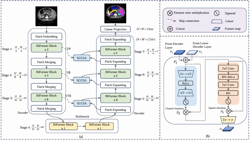
BRAU-Net++: The Hybrid CNN-Transformer That Rethinks Sparse Attention for Medical Image Segmentation.

BRAU-Net++: U-Shaped Hybrid CNN-Transformer Network for Medical Image Segmentation

Researchers at Chongqing University of Technology built a u-shaped encoder-decoder that fuses dynamic sparse attention from BiFormer with a redesigned channel-spatial skip connection — outperforming TransUNet by 4.49% DSC and Swin-Unet by 3.34% DSC on the Synapse multi-organ benchmark while…

BRAU-Net++: U-Shaped Hybrid CNN-Transformer Network for Medical Image Segmentation Read More »

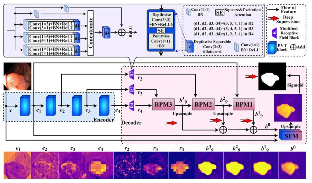
MSBP-Net: The Lightweight Polyp Detector.

MSBP-Net: The Lightweight Polyp Detector That Learned to See Boundaries the Way Surgeons Do

Researchers at Sichuan University of Science and Engineering built a network that fuses reverse attention, multi-scale criss-cross self-attention, and a Laplacian edge filter borrowed from classical image processing to delineate colorectal polyp boundaries with a precision and speed that most…

MSBP-Net: The Lightweight Polyp Detector That Learned to See Boundaries the Way Surgeons Do Read More »

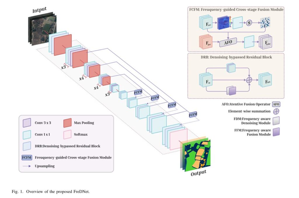
Overview of the proposed FreDNet.

FreDNet: The Remote Sensing Segmenter That Learned to Hear the Image, Not Just See It

Researchers at Hohai University built a network that treats high-frequency spectral components — the sharp edges, fine contours, and subtle discontinuities that standard convolutions quietly destroy — as first-class semantic cues rather than noise to be filtered away, and the…

FreDNet: The Remote Sensing Segmenter That Learned to Hear the Image, Not Just See It Read More »

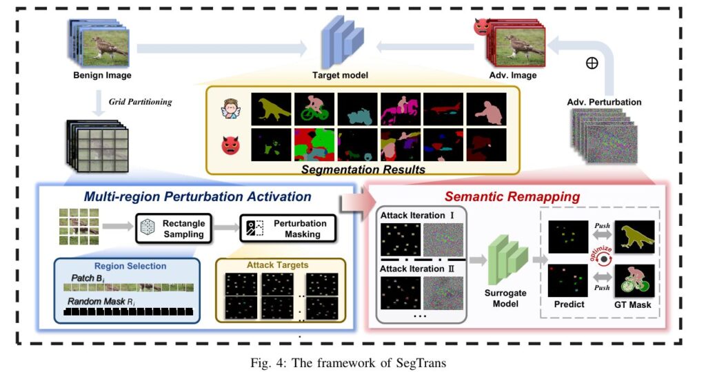
The framework of SegTrans.

SegTrans: The Transfer Attack That Finally Broke Segmentation Models (Without Extra Compute)

Segmentation models correct each other’s mistakes through a “tight coupling” phenomenon – which makes them brutally hard to fool in a black‑box setting. Researchers discovered that by destroying global semantic integrity and remapping local features, transfer attack success jumps by…

SegTrans: The Transfer Attack That Finally Broke Segmentation Models (Without Extra Compute) Read More »

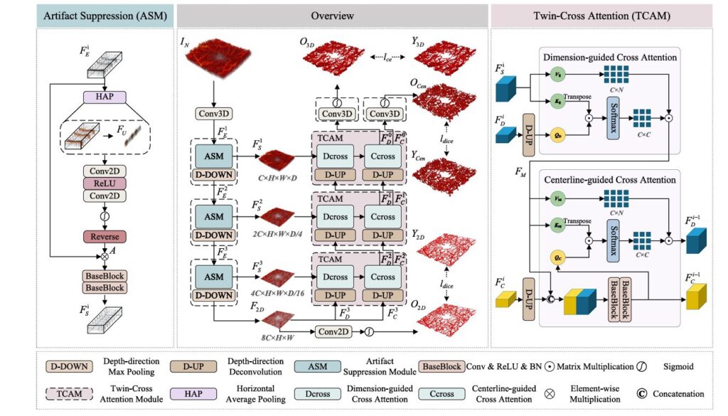
The MT-Net encoder-decoder architecture with dimension transformation. D-DOWN operations compress depth while preserving lateral structure; D-UP operations restore volumetric resolution during decoding

MT-Net: 3D Retinal Microvascular Segmentation via Multi-Scale Topology Regulation

Optical coherence tomography angiography arrived in clinical practice with a peculiar contradiction. It captures volumetric blood flow at micron resolution, yet the vast majority of analysis tools treat it as a stack of 2D images. The enface projection — a…

MT-Net: 3D Retinal Microvascular Segmentation via Multi-Scale Topology Regulation Read More »

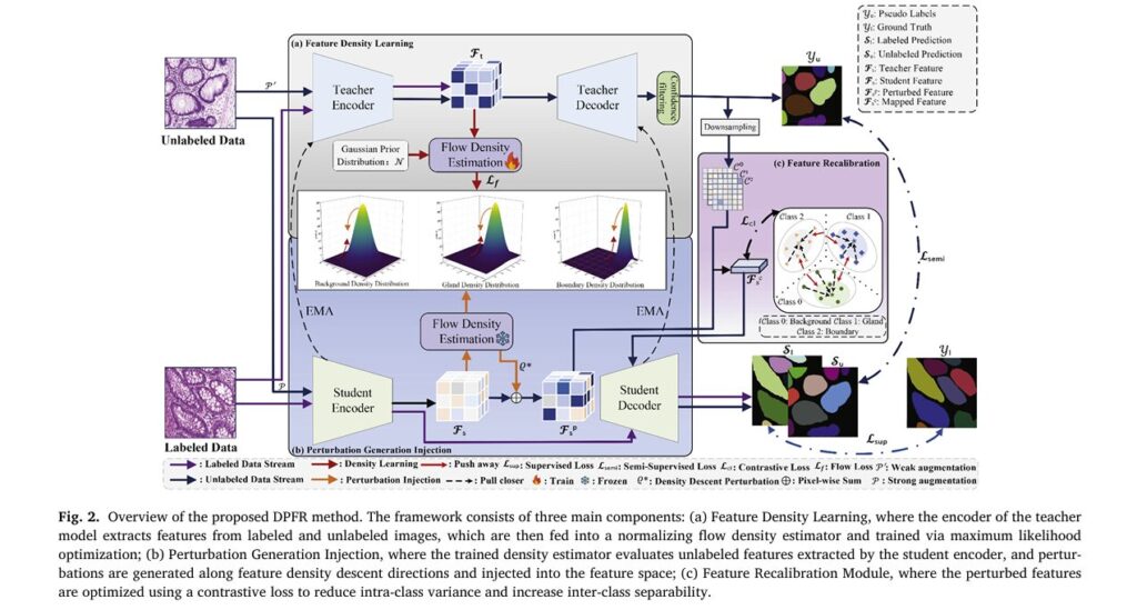
DPFR: A Breakthrough in AI-Powered Gland Segmentation for Cancer Diagnosis

DPFR: A Breakthrough in AI-Powered Gland Segmentation for Cancer Diagnosis

The early detection and accurate grading of cancer remains one of modern medicine’s most pressing challenges. For pathologists worldwide, the assessment of gland morphology in histopathological images serves as the gold standard for cancer diagnosis—particularly in colorectal and prostate cancers.…

DPFR: A Breakthrough in AI-Powered Gland Segmentation for Cancer Diagnosis Read More »