Image Segmentation

Image segmentation is the task of labeling an image at the pixel level, and it underpins much of modern medical imaging, autonomous perception, and scene understanding. Here we break down segmentation architectures, loss functions, and evaluation practices, with frequent attention to the medical settings where boundary accuracy and robustness matter most. Each piece traces the method back to its source research.

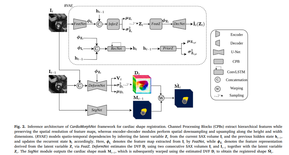
CardioMorphNet: Shape-Guided Bayesian Recurrent Deep Learning for 3D Cardiac Motion Estimation.

CardioMorphNet: Shape-Guided Bayesian Recurrent Deep Learning for 3D Cardiac Motion Estimation

Researchers at the University of Glasgow and the University of Manchester built a shape-guided Bayesian recurrent framework that estimates 3D cardiac motion from cine CMR images without relying on intensity-based image registration — and it outperforms every major method on…

CardioMorphNet: Shape-Guided Bayesian Recurrent Deep Learning for 3D Cardiac Motion Estimation Read More »

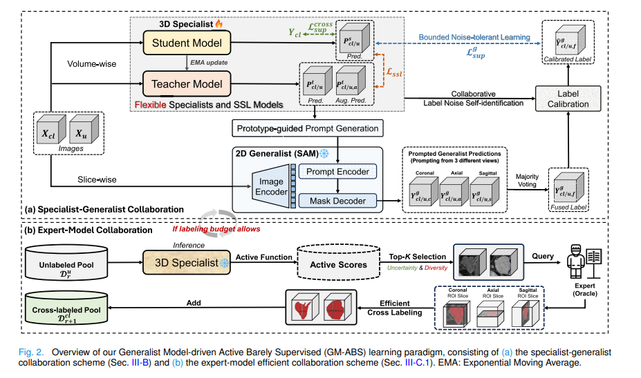
GM-ABS: SAM-Driven Active Barely Supervised 3D Medical Image Segmentation.

GM-ABS: SAM-Driven Active Barely Supervised 3D Medical Image Segmentation

Researchers from CUHK and Harvard Medical School built a training paradigm where the in-training specialist autonomously crafts prompts for a frozen SAM generalist, which returns noisy-but-useful labels across three orthogonal views — while active learning selects the most informative scans…

GM-ABS: SAM-Driven Active Barely Supervised 3D Medical Image Segmentation Read More »