
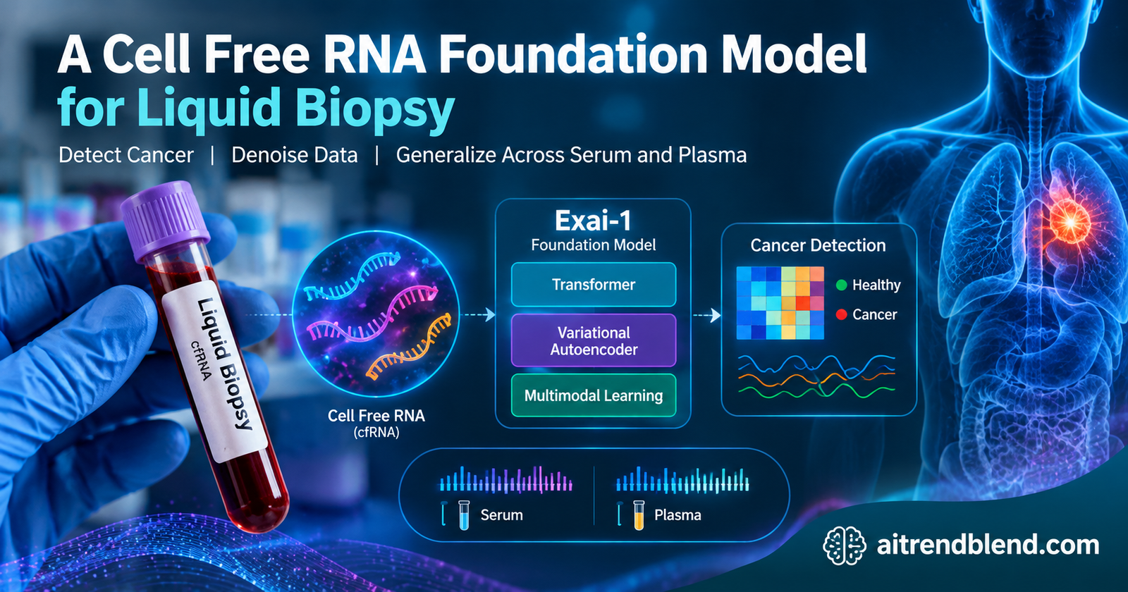
A tube of blood holds a faint rumour of what is happening deep inside the body. Among the debris floating in plasma are fragments of RNA shed by cells, and when a tumour is growing somewhere, it leaks its own telltale fragments into that stream. Reading them could catch a cancer early, from a routine blood draw, with no biopsy needle.
The trouble is that the signal is faint, scattered, and easily drowned out by noise. A team at Exai Bio, working with three universities, built a foundation model to pull that signal out, and named it Exai-1.
Key points
- Exai-1 is a foundation model for cell free RNA, the RNA fragments that circulate in blood, built to make blood based cancer detection more reliable.
- It combines two views of each RNA fragment, what its sequence means and how abundant it is, inside a transformer paired with a variational autoencoder, and was pretrained on more than 300 billion tokens from over 8,000 blood samples.
- It reconstructs noisy or missing measurements with high fidelity, reaching a fit far above the usual baseline, and it cleans up noisy data better than a standard imputation method.
- Its learned representation lets a cancer classifier trained on plasma still work on serum, a translation that failed completely when the classifier used raw measurements.
- Synthetic blood profiles it generates can pad out small datasets and lift cancer detection accuracy, which matters most for rare cancers where real samples are scarce.
The signal hiding in a blood draw
Liquid biopsy is the idea that you can learn about a disease inside the body by reading molecules that leak into the blood, without cutting anything out. Most attention has gone to circulating tumour DNA, the genetic fragments a dying cancer cell releases. This paper works with something different and, the authors argue, richer. Cell free RNA, or cfRNA, is a soup of RNA fragments circulating in blood, and a particular class of them turns out to carry a strong cancer signal.
The team had previously identified what they call orphan non coding RNAs, small cancer emergent RNA fragments that healthy cells barely produce but tumours actively secrete into the blood, often at higher levels than tumour DNA. The vivid image the authors use is a constellation. Any single one of these fragments means little on its own, the way one star is just a dot, but their collective pattern spells out the presence and even the type of a cancer. Reading that pattern is exactly the kind of problem where machine learning should shine.
What stops it from being easy is the data. Blood samples are precious and often scarce, especially for rare cancers, and the measurements are high dimensional and noisy, with thousands of RNA features measured across only a few thousand samples. Worse, the numbers shift depending on which biofluid was used, serum or plasma, and which lab processed the sample, so a model trained in one setting often falls apart in another. That combination of scarcity, noise, and inconsistency is the wall Exai-1 is built to climb. The broader move toward reading disease from molecular data runs through our coverage of AI for early disease detection.
Two views of every RNA fragment
The core idea is to look at each RNA fragment from two angles at once. One angle is meaning. Every fragment has a sequence, a string of RNA letters, and that sequence carries clues about what the fragment does and where it came from. The other angle is quantity, simply how much of that fragment is present in a given sample. Older methods tended to use one or the other. Exai-1 fuses them, so the model knows both what a fragment is and how loud its signal is in this particular blood draw.
The meaning half comes from a separate RNA foundation model called RNA-FM, which has already learned rich representations of RNA sequences. Exai-1 uses those learned sequence embeddings to initialize its own understanding of each fragment, then scales each one by how abundant the fragment is in the sample. Reading a blood sample becomes something like reading a sentence, where each RNA fragment is a word carrying both a meaning and an emphasis. That sentence style framing of biology echoes our look at generative modelling of RNA structure, which reads the molecule from a different angle.
Exai-1 treats a blood sample as a sentence of RNA fragments, each carrying both a learned meaning from its sequence and an emphasis from its abundance. Fusing the two is what lets the model find biological structure that either view alone would miss.
Learning by filling in the gaps
The model learns the way many language models do, by hiding part of the input and learning to fill it back in. During training Exai-1 masks a quarter of the RNA measurements in each sample and tries to reconstruct them from the rest, which forces it to learn the dependencies among fragments rather than memorizing any single one. That is the same self supervised trick that powers text models, applied to the vocabulary of circulating RNA.
Around that reconstruction sit two more ideas that give the model its character. The first is a variational autoencoder, which squeezes each sample through a narrow probabilistic bottleneck of just thirty two numbers before reconstructing it. That bottleneck forces the model to keep only what matters and discard noise, and because it is probabilistic, the model can later sample from it to generate new, realistic blood profiles.
The second idea is how the model handles the pesky technical differences between labs and biofluids. Rather than blindly erasing every difference between samples, which risks scrubbing out real biology, Exai-1 uses a triplet margin loss guided by known biological labels. It pulls samples that share a true biological trait, such as having the same cancer, closer together in its latent space, and pushes samples with different traits apart, using the biology to decide what should be similar rather than letting lab artefacts drive the grouping.
All of this trains together under one combined objective that adds up the reconstruction of the visible and masked features, the variational term, the biological grouping term, and a set of prediction tasks for cancer status, tissue of origin, biofluid type, and assay version. Those last tasks are carried by special tokens the model always reads, a design that lets it fold clinically relevant labels directly into its representation.
A notable point is how small the model is. Because it works on a compressed representation rather than the full high dimensional input, Exai-1 has only about 3.6 million trainable parameters and trained on a single modest graphics card, which is tiny by the standards of modern foundation models and a deliberate guard against overfitting on limited data.
What the results show
The paper builds its case in layers, starting with the most basic question of whether the model reconstructs data faithfully and ending with whether it actually helps detect cancer.
| Task | What was measured | Exai-1 | Comparison |
|---|---|---|---|
| Reconstructing masked measurements | Goodness of fit | 0.89 | 0.57 for the average baseline |
| Cancer detection on serum, trained on plasma | Area under the curve | 0.74 using the latent space | 0.56 using raw measurements |
| Ovarian cancer, a small cohort | Gain in area under the curve | Up 0.10 from the latent space | Over raw measurements |
| Batch effect correction | Aggregate benchmark score | 0.34 | 0.28 for the next best method |
The reconstruction result is the foundation. Asked to recover measurements it had never seen, Exai-1 reached a fit far above the baseline that many models struggle to beat, and it kept the biological structure intact, preserving the clusters of related RNA fragments rather than smoothing them into mush. When the team deliberately added noise at eight increasing levels, the model kept cleaning it up effectively, outperforming a standard graph based imputation method that fell apart as the noise grew.
The most clinically pointed result is about generalizing across biofluids. A cancer classifier trained on plasma and then tested on serum, using the raw RNA measurements, was barely better than a coin flip. The same classifier, trained on Exai-1’s learned representation instead, held up on serum almost as well as on plasma. By separating the biological signal from the technical fingerprint of how a sample was collected, the model let a classifier built in one setting survive in another, which is a real obstacle in deploying these tests.
Exai-1 decouples the biological signal from technical variations within the latent space, making it possible for a plasma trained model to generalize to out of distribution serum data, a task that was not possible in the original feature space. Karimzadeh and colleagues, Nature Machine Intelligence, 2025
The generative angle pays off where data is thinnest. Because the model can dream up realistic synthetic blood profiles, the team used it to pad out small training sets and found the cancer detection accuracy improved, with the biggest gains on the smallest datasets. On a small ovarian cancer cohort, moving from raw measurements to the model’s representation lifted the detection score meaningfully, and adding synthetic samples nudged it up further. For rare cancers where you may only have a few dozen samples, that kind of augmentation is exactly the lever you want. The same appetite for squeezing more signal from limited biological data runs through our analysis of fusing pathology images with gene expression to predict cancer outcomes.
Reading what the model learned
A recurring worry with foundation models is that they become inscrutable, so the authors spend real effort showing that Exai-1’s internal representation lines up with biology. Its embedding dimensions turned out to correspond to genuine groups of RNA fragments that rise and fall together, and when they traced which fragments most drove the model’s cancer predictions, the orphan RNAs stood out, with nearly three quarters of them contributing strongly, more than any other class of fragment.
Those influential fragments mapped onto genes and processes already tied to cancer, including known cancer associated loci and biological pathways for cell division, programmed cell death, and cell movement, the very machinery that goes wrong in a tumour. A model that not only predicts but points back at plausible cancer biology is far more trustworthy than a black box, and it hints at the same fragments being useful as biomarkers in their own right.
From a research model to a clinical test
This is where enthusiasm has to give way to care, because the distance between an impressive benchmark and a test a doctor can order is long, and a few things about this study make that distance worth stating plainly.
The results come from a large but retrospective dataset, samples already collected and labelled, evaluated on a held out portion of that same collection. That is the right way to develop a model, and it is not the same as a prospective clinical trial, where the test is run on new patients and judged against what actually happens to them. The authors are explicit that clinical validation on independent datasets is a necessary next step, and until that is done the reported detection scores describe performance on a research benchmark, not a screening test’s real world accuracy.
The numbers themselves, read honestly, are a research signal rather than a finished diagnostic. A detection score around three quarters is a meaningful advance for a hard problem, and it is not the near certainty a population screening test would demand, where a false alarm sends a healthy person into anxious follow up and a miss provides false reassurance. Cancer detection from blood also has to clear a high bar on exactly the cases that are hardest and matter most, the small, early tumours that shed the least into the blood, and a retrospective benchmark skewed toward samples with a known diagnosis does not fully probe that regime.
One more thing belongs in an honest read. Several of the authors, including senior ones, are founders, shareholders, or advisors of the company commercializing this technology, which they disclose. That does not make the science wrong, and the work is peer reviewed and its code and data released, but a diagnostic claim from a team with a direct commercial stake is precisely the kind that independent, prospective validation exists to check. The paper presents a foundation for a test, not a cleared test.
Exai-1 is a strong research foundation for blood based cancer detection, not a validated clinical test. Its scores come from a retrospective benchmark, the hardest early stage cases remain the real challenge, and the commercial interests behind it are exactly why independent prospective validation matters before anyone relies on it.
Where the method falls short
Beyond the clinical gap, the authors and a careful reading surface several technical limits.
The whole approach rests on the orphan RNAs the same group discovered, and while the model treats them as a rich signal, the biology of why tumours secrete them and how consistently they appear across patients and cancer types is still being worked out. A representation is only as good as the signal underneath it, and if those fragments behave differently in populations unlike the training cohort, the model’s advantage could narrow.
The synthetic data that helps small cohorts is a double edged tool. The paper is candid that augmentation occasionally hurt rather than helped, particularly when reconstructions were generated from very small or biased training sets, or when the baseline dataset was already large enough that added variability just reduced the signal to noise ratio. Generated samples inherit whatever the model got wrong, so leaning on them is safest exactly where it is also riskiest, in the data starved corners.
There are quieter caveats too. The model was trained and evaluated entirely on data from a single processing pipeline, diverse across suppliers and sites but still one methodology, so how it behaves on samples processed differently is untested. And like any model that compresses to a tiny latent space, it makes a bet that thirty two numbers can hold what matters, a bet that works well here but could lose fine detail that turns out to be important for a subtler task than the ones benchmarked.
Why it matters
Step back and the contribution is a way to make a fragile kind of data usable. Cell free RNA carries a real cancer signal, but scarcity, noise, and inconsistency between labs had kept it from reaching its potential. By fusing sequence meaning with abundance, compressing to a denoised representation, and correcting technical differences with biology rather than blind erasure, Exai-1 turns a temperamental measurement into something a downstream classifier can rely on and even generalize from.
The broader significance is the template. A model that pretrains on abundant, imperfect molecular data and transfers to scarce clinical settings is chasing the same goal across biomedicine, extracting more from every precious patient sample. The specific tricks here, a biologically guided batch correction and generative augmentation for rare cases, are not tied to cfRNA and could travel to other molecular measurements that suffer the same scarcity. As foundation models spread from language into the messy signals of the body, from the drug response models to the imaging tools in our wider coverage of AI in drug discovery and biology, the ones that stay small, interpretable, and honest about the clinical gap are the ones worth building on.
A reference implementation
The code below is a compact, runnable version of the core architecture. It builds a multimodal variational autoencoder with a self attention encoder over RNA tokens, a probabilistic bottleneck, a self attention decoder that reconstructs masked measurements, and the combined objective of reconstruction, the variational term, and a triplet margin loss for biological grouping, with a smoke test on dummy data. The authors full code and models are linked under the block.
# Exai-1 style multimodal cfRNA foundation model, compact reference
# A self attention variational autoencoder reconstructs masked RNA measurements
# and a triplet loss groups samples by biology, not by lab batch.

import torch
import torch.nn as nn
import torch.nn.functional as F

torch.manual_seed(0)


class SelfAttnBlock(nn.Module):
    def __init__(self, dim, heads=4):
        super().__init__()
        self.attn = nn.MultiheadAttention(dim, heads, batch_first=True)
        self.norm1, self.norm2 = nn.LayerNorm(dim), nn.LayerNorm(dim)
        self.ff = nn.Sequential(nn.Linear(dim, dim * 2), nn.GELU(), nn.Linear(dim * 2, dim))

    def forward(self, x):
        a, _ = self.attn(x, x, x)
        x = self.norm1(x + a)
        return self.norm2(x + self.ff(x))


class ExaiVAE(nn.Module):
    def __init__(self, n_features, seq_dim=32, latent=32):
        super().__init__()
        # each RNA fragment carries a sequence embedding scaled by its abundance
        self.seq_embed = nn.Embedding(n_features, seq_dim)
        self.enc = SelfAttnBlock(seq_dim)
        self.to_mu = nn.Linear(n_features * seq_dim, latent)
        self.to_logvar = nn.Linear(n_features * seq_dim, latent)
        self.dec_in = nn.Linear(latent, n_features * seq_dim)
        self.dec = SelfAttnBlock(seq_dim)
        self.readout = nn.Linear(seq_dim, 1)
        self.n_features, self.seq_dim = n_features, seq_dim

    def encode(self, abundance, ids):
        # signal embedding, sequence meaning times abundance
        tok = self.seq_embed(ids) * abundance.unsqueeze(-1)
        h = self.enc(tok)
        flat = h.flatten(1)
        return self.to_mu(flat), self.to_logvar(flat)

    def reparam(self, mu, logvar):
        std = (0.5 * logvar).exp()
        return mu + std * torch.randn_like(std)     # sample the latent

    def decode(self, z):
        b = z.shape[0]
        h = self.dec_in(z).view(b, self.n_features, self.seq_dim)
        h = self.dec(h)
        return self.readout(h).squeeze(-1)          # reconstructed abundances

    def forward(self, abundance, ids):
        mu, logvar = self.encode(abundance, ids)
        z = self.reparam(mu, logvar)
        return self.decode(z), mu, logvar, z


def kl_term(mu, logvar):
    return -0.5 * torch.mean(1 + logvar - mu ** 2 - logvar.exp())


def triplet_bio_loss(z, labels, margin=0.1):
    # pull same label samples together, push different label samples apart
    d = torch.cdist(z, z)
    loss, count = 0.0, 0
    for i in range(len(labels)):
        pos = (labels == labels[i]) & (torch.arange(len(labels)) != i)
        neg = labels != labels[i]
        if pos.any() and neg.any():
            loss = loss + F.relu(d[i][pos].mean() - d[i][neg].mean() + margin)
            count += 1
    return loss / max(count, 1)


if __name__ == "__main__":
    n_features, batch = 200, 16
    model = ExaiVAE(n_features)
    opt = torch.optim.Adam(model.parameters(), lr=1e-3)

    ids = torch.arange(n_features).unsqueeze(0).expand(batch, -1)
    abundance = torch.rand(batch, n_features)               # normalized RNA levels
    labels = torch.randint(0, 2, (batch,))                  # cancer or control

    for step in range(200):
        mask = (torch.rand(batch, n_features) > 0.25).float()   # hide 25 percent
        recon, mu, logvar, z = model(abundance * mask, ids)
        recon_loss = F.smooth_l1_loss(recon, abundance)
        loss = 100 * recon_loss + 10 * kl_term(mu, logvar) + triplet_bio_loss(z, labels)
        opt.zero_grad()
        loss.backward()
        opt.step()
        if (step + 1) % 50 == 0:
            print(f"step {step + 1}  loss {loss.item():.4f}  recon {recon_loss.item():.4f}")
Go to the source
Read the peer reviewed paper and explore the authors open code and data.
Read the paper Code on GitHubConclusion
The core achievement here is a foundation model that makes cell free RNA usable. By reading each circulating fragment through two lenses at once, what its sequence means and how abundant it is, and by compressing thousands of noisy measurements into a small denoised representation, Exai-1 turns a fragile and inconsistent signal into something a cancer classifier can rely on. It reconstructed missing measurements far better than the usual baseline, cleaned up noise where a standard method faltered, and let a classifier trained on one biofluid survive on another.
The conceptual shift worth remembering is the choice to correct technical differences with biology rather than against it. Instead of blindly erasing every difference between samples, which risks scrubbing away the signal, the model uses known biological labels to decide what should look alike, and pairs that with a generative bottleneck that can invent new realistic samples where real ones are scarce. Those two ideas, biologically guided batch correction and generative augmentation, are the engine behind its gains on rare cancers.
The approach travels because its problem is everywhere in biomedicine. Scarce, noisy, inconsistent molecular data from precious patient samples is the norm, not the exception, and a small, interpretable model that pretrains on abundant imperfect data and transfers to thin clinical settings is a template well beyond cfRNA. That the model stays tiny and lets its influential features be traced back to real cancer biology only strengthens the case.
The honest limits keep it grounded. The results are retrospective rather than from a prospective trial, the detection scores are a research signal rather than a screening test’s accuracy, the hardest early stage cancers remain the real challenge, augmentation can hurt as well as help, and the commercial interests behind the work are exactly why independent validation matters. This is a foundation for a test, not a test.
Future directions follow naturally. Validate the model prospectively on new patients and independent cohorts, test it on samples processed through other pipelines, and push toward the early, low signal tumours where blood based detection would change the most lives. If those steps hold up, the faint rumour in a tube of blood could become something a clinic can actually read, and the promise of catching cancer early from a routine draw would move a real step closer.
Frequently asked questions
What is Exai-1?
Exai-1 is a foundation model for cell free RNA, the RNA fragments that circulate in blood. It combines each fragment’s sequence meaning with how abundant it is, and was pretrained on billions of tokens from over 8,000 blood samples to support blood based cancer detection.
What is cell free RNA and why does it matter for cancer?
Cell free RNA is a mix of RNA fragments floating in the blood. A class of them called orphan RNAs is barely produced by healthy cells but is actively released by tumours, so their collective pattern can signal the presence and type of a cancer from a simple blood draw.
How well does the model actually work?
On a research benchmark it reconstructed missing measurements with a fit of about 0.89 against a baseline of 0.57, and its representation let a cancer classifier trained on plasma still reach an area under the curve of 0.74 on serum, where raw measurements managed only 0.56. These are research results, not a validated clinical test.
Can this detect cancer from my blood today?
No. Exai-1 is a research foundation model evaluated on a retrospective dataset, not a cleared diagnostic test. Turning it into something a doctor could order would require prospective clinical trials on new patients and independent validation, which the authors state is a necessary next step.
Why does the model generate synthetic blood samples?
Its variational design can sample new, realistic blood profiles, which are used to pad out small training sets. This helped most for rare cancers where only a few dozen real samples exist, though the authors note that augmentation can sometimes hurt when the base data is very small or biased.
Can the code and data be reproduced?
Yes. The authors released the Exai-1 code on GitHub and Zenodo and the pretrained model and expression matrices on Zenodo, with other data available from the authors under data use agreements, so the key results can be reproduced.