Image Segmentation

Image segmentation is the task of labeling an image at the pixel level, and it underpins much of modern medical imaging, autonomous perception, and scene understanding. Here we break down segmentation architectures, loss functions, and evaluation practices, with frequent attention to the medical settings where boundary accuracy and robustness matter most. Each piece traces the method back to its source research.

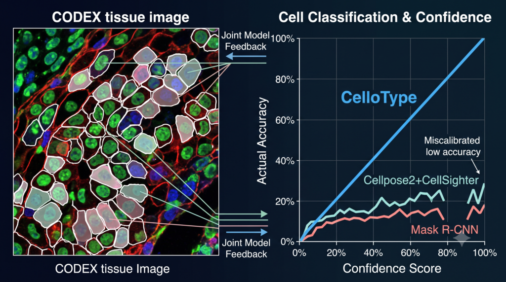
A Single Model for Cell Segmentation and Classification Finally Gets Confidence Scores Right

Analysis by the aitrendblend editorial team · Source paper doi.org/10.1038/s41592-024-02513-1 Spatial Omics Cell Segmentation Transformers Multitask Learning Nature Methods A CODEX tissue image with cells outlined by CelloType and each one tagged with a predicted type and a confidence percentage. Every spatial omics pipeline starts the same way. Find the cells, then figure out what […]

A Single Model for Cell Segmentation and Classification Finally Gets Confidence Scores Right Read More »

MedDINOv3: Revolutionizing Medical Image Segmentation with Adaptable Vision Foundation Models

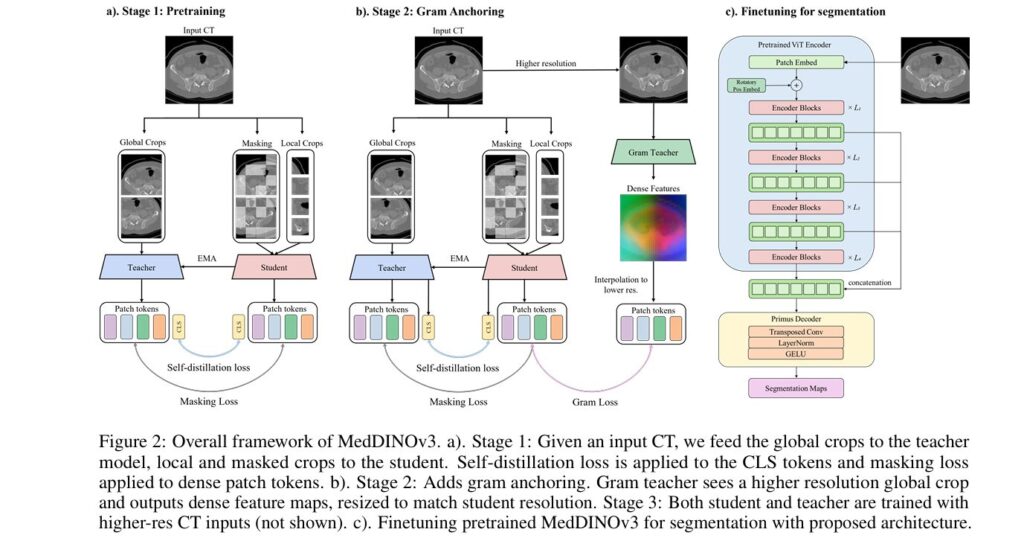
MedDINOv3 Adapts A Vision Foundation Model For CT And MRI Segmentation

AI for medical imaging and healthcare Vision foundation models CT and MRI segmentation Self supervised pretraining Analysis by the aitrendblend editorial team A radiation oncologist planning a course of treatment needs the kidneys, liver, spinal cord, and every nearby organ outlined precisely enough that the radiation beam avoids them by design rather than by luck.

MedDINOv3 Adapts A Vision Foundation Model For CT And MRI Segmentation Read More »

Balancing Conflict Gradients in Semi Supervised Segmentation

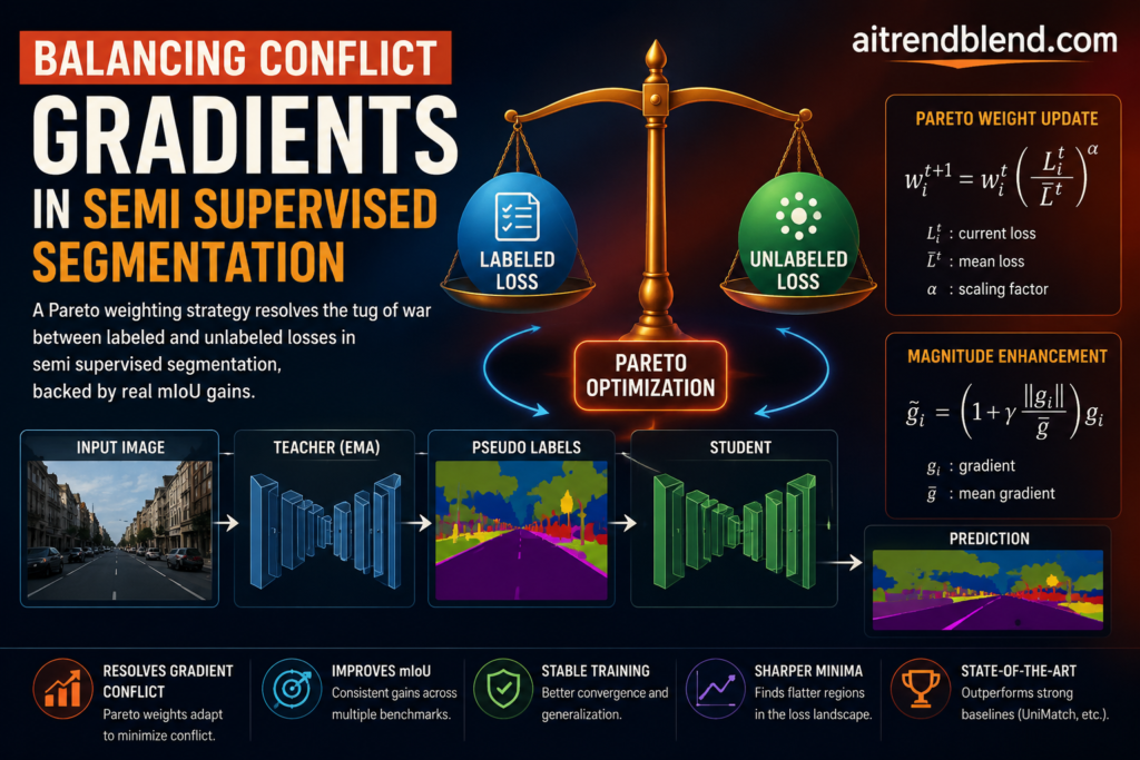
Balancing Conflict Gradients in Semi Supervised Segmentation

Analysis by the aitrendblend editorial team · 12 min read · Semi supervised learning and training strategies semi supervised segmentation gradient conflict Pareto optimization teacher student networks UniMatch Supervised and unsupervised gradients often point in different directions during training. A Pareto weighting scheme finds the direction that helps both at once. Somewhere around the four

Balancing Conflict Gradients in Semi Supervised Segmentation Read More »

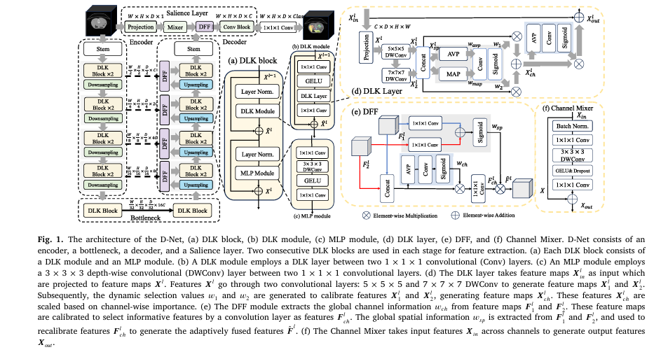
D-Net: A New Frontier in AI-Powered Medical Image Segmentation

D-Net Pairs Dynamic Large Kernels With a Pixel Level Salience Layer

Medical Imaging Biomedical Signal Processing and Control, Volume 113, 2026 23 minute read Volumetric Segmentation Dynamic Large Kernel Vision Transformer CT and MRI Organ Segmentation Tumor Segmentation Feature Fusion Receptive Field D-Net This article describes a peer reviewed computer science paper about an automatic image segmentation research tool. It reports benchmark results on public research

D-Net Pairs Dynamic Large Kernels With a Pixel Level Salience Layer Read More »

GRCSF’s Dual-Feature Compensation Framework Achieves State-of-the-Art Lesion Segmentation

How GRCSF Compensates for Lost Detail in Medical Lesion Segmentation

Analysis by the aitrendblend editorial team. Source paper, Wang, Chen, Yang and Kim, Pattern Recognition, 2026. lesion segmentation stroke imaging lung tumor CT coronary calcium scoring self supervised learning masked autoencoders A radiologist studying a T1 weighted brain scan after a stroke is often hunting for a patch of tissue that looks almost exactly like

How GRCSF Compensates for Lost Detail in Medical Lesion Segmentation Read More »

U-Mamba2-SSL: The Groundbreaking AI Framework Revolutionizing Tooth & Pulp Segmentation in CBCT Scans

U-Mamba2-SSL Segments Teeth and Pulp From Unlabeled CBCT

Analysis by the aitrendblend editorial team, based on the published paper and an independent read of its claims. Not a substitute for advice from a licensed dentist or clinician. Medical Imaging AI CBCT Segmentation Semi Supervised Learning Dental AI State Space Models Picture a hospital archive full of cone beam CT scans of people’s jaws,

U-Mamba2-SSL Segments Teeth and Pulp From Unlabeled CBCT Read More »

CMFDNet Tackles Blurry Polyp Boundaries With A Cross Mamba Decoder

CMFDNet Tackles Blurry Polyp Boundaries With A Cross Mamba Decoder

AI for medical imaging and healthcare Polyp segmentation Mamba architectures Colonoscopy AI Analysis by the aitrendblend editorial team A four stage encoder feeds a cross scanning Mamba decoder that fuses deep and shallow polyp features before a final feature discovery pass. A gastroenterologist pulling a colonoscope back through the colon has maybe a second or

CMFDNet Tackles Blurry Polyp Boundaries With A Cross Mamba Decoder Read More »

Towards Trustworthy Breast Tumor Segmentation in Ultrasound Using AI Uncertainty

Analysis by the aitrendblend editorial team · Source paper arXiv:2508.17768 Medical Imaging Segmentation Uncertainty Estimation Breast Ultrasound nnU-Net An ultrasound frame next to the kind of entropy map the model produces when it is asked to also grade its own confidence. A radiologist scanning a breast for a suspicious mass rarely gets a clean answer

Towards Trustworthy Breast Tumor Segmentation in Ultrasound Using AI Uncertainty Read More »

RoofSeg: An edge-aware transformer-based network for precise roof plane segmentation from LiDAR point clouds

RoofSeg Explained, End to End Roof Plane Segmentation From LiDAR

COMPUTER VISION & GEOSPATIAL AI · 14 MIN READ · Analysis by the aitrendblend editorial team RoofSeg airborne LiDAR roof plane segmentation edge-aware transformer PointNet++ 3D building reconstruction Turning a scatter of LiDAR points into a clean 3D model of a building roof sounds like a job for careful geometry, and for a long time

RoofSeg Explained, End to End Roof Plane Segmentation From LiDAR Read More »

Task-Specific Knowledge Distillation in Medical Imaging: A Breakthrough for Efficient Segmentation.

Task-Specific Knowledge Distillation for Medical Image Segmentation

Knowledge Distillation Medical Image Segmentation • 15 min read Task-Specific KD Segment Anything LoRA ViT-Tiny Diffusion Data Data-Limited Learning Teaching a Tiny Model to Segment Like a Giant Overview. A large vision foundation model is first adapted to one medical task with LoRA, then it teaches a compact student through both its hidden features and

Task-Specific Knowledge Distillation for Medical Image Segmentation Read More »