Image Segmentation

Image segmentation is the task of labeling an image at the pixel level, and it underpins much of modern medical imaging, autonomous perception, and scene understanding. Here we break down segmentation architectures, loss functions, and evaluation practices, with frequent attention to the medical settings where boundary accuracy and robustness matter most. Each piece traces the method back to its source research.

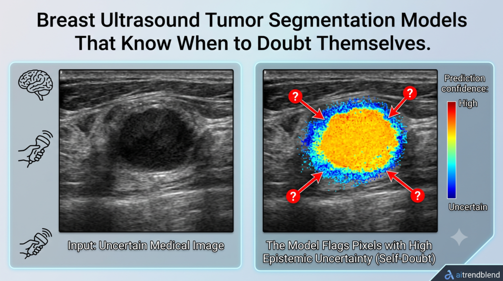
Towards Trustworthy Breast Tumor Segmentation in Ultrasound Using AI Uncertainty

This article explains a published research paper. It is not medical advice, diagnosis, or treatment guidance. The segmentation model discussed here is a research prototype, not an approved clinical device. Anyone with a health concern about a breast finding should…

Towards Trustworthy Breast Tumor Segmentation in Ultrasound Using AI Uncertainty Read More »

Proposed DSCA NET for image Segmentation

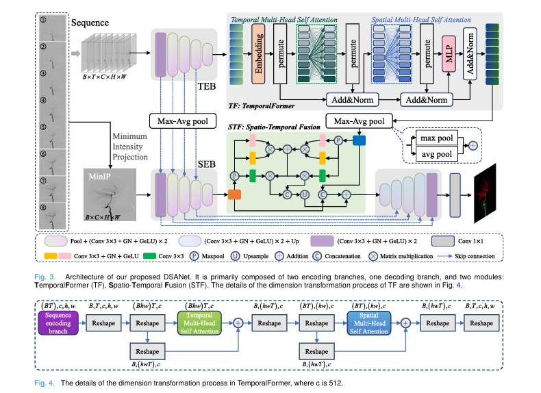
DSANET Uses Full DSA Sequences to Segment Cerebral Arteries

Cerebrovascular diseases, stroke, moyamoya disease, and cerebral aneurysms among them, remain a major cause of death and disability, and diagnosing them accurately depends on understanding the geometry of the cerebral arteries. Digital subtraction angiography, often shortened to DSA, is the…

DSANET Uses Full DSA Sequences to Segment Cerebral Arteries Read More »

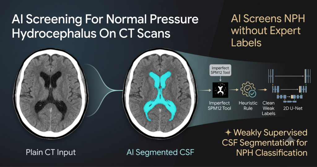
Weakly Supervised AI for Normal Pressure Hydrocephalus (NPH) Screening on CT Scans

Weakly Supervised AI for Normal Pressure Hydrocephalus (NPH) Screening on CT Scans

Normal pressure hydrocephalus, usually shortened to NPH, shows up mostly in older adults as a triad of gait impairment, urinary incontinence, and cognitive decline, caused by cerebrospinal fluid building up in the brain’s ventricular system. What makes it clinically important,…

Weakly Supervised AI for Normal Pressure Hydrocephalus (NPH) Screening on CT Scans Read More »